At a Glance
The trachea (windpipe) is the main airway between the larynx and the lungs. In adults it is about 10 to 12 cm long and about 2 cm wide, held open by 16 to 20 C-shaped rings of hyaline cartilage. At the level of the sternal angle it splits into the right and left main bronchi.
- Location
- Midline of the lower neck and upper chest, from the larynx (C6 level) to the sternal angle (T4-T5 level)
- Function
- Conducts air to and from the lungs, warms, moistens and cleans it, and triggers coughing
- Tissue
- Ciliated pseudostratified columnar epithelium, submucosal glands, C-shaped hyaline cartilage rings, trachealis smooth muscle
- Size
- About 10-12 cm long and about 2 cm in diameter in adults
- Connects to
- Larynx above; right and left main bronchi below, at the carina
- Part of
- Lower respiratory tract (conducting zone)
On this page
- What Is the Trachea?
- Where Is the Trachea Located in the Body?
- Trachea Anatomy: Cartilage Rings, Trachealis Muscle and Carina
- What Is the Trachea Made Of?
- What Does the Trachea Do?
- How Does the Trachea Work During Breathing?
- Trachea vs Esophagus: What Is the Difference?
- How Does the Trachea Develop and Change With Age?
- Common Conditions That Affect the Trachea
- Key Facts
- Quiz
- FAQ
What Is the Trachea?
The trachea is the medical name for the windpipe: a stiff but flexible tube that links the larynx (voice box) to the two main bronchi and carries every breath into and out of the lungs. It is the widest airway below the throat and the first part of the lower respiratory tract, which continues through the bronchi and bronchioles to the alveoli. Above it lies the upper respiratory tract: the nose, nasal cavity, pharynx and larynx.
What does the trachea look like?
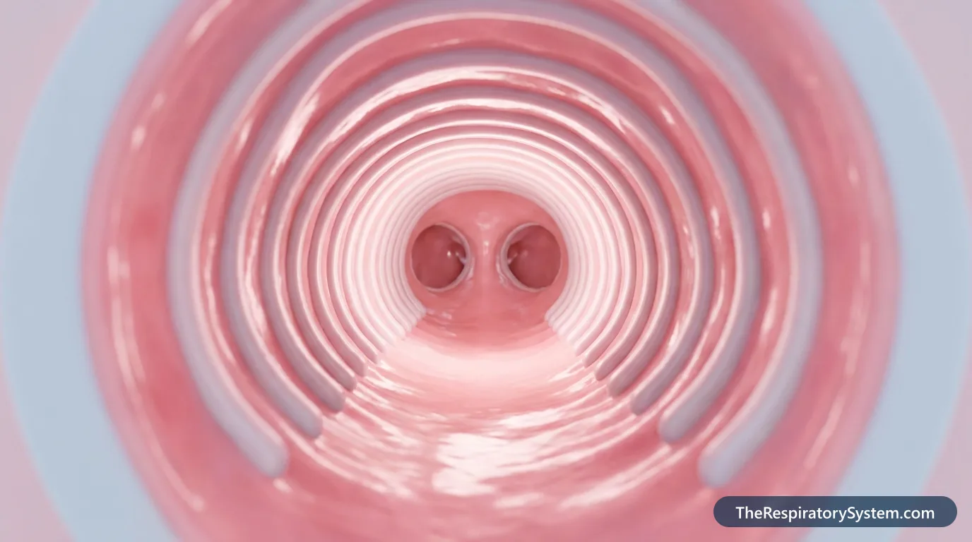
From the outside, the trachea looks like a short length of ribbed vacuum hose. A stack of pale, horseshoe-shaped cartilage rings gives it its ridged surface, and narrow bands of fibrous tissue join one ring to the next. The back of the tube is flat and soft, because the rings are open at the back and the gap is closed by muscle and membrane. Inside, the lining is pink and glistening with a thin coat of mucus.
How big is the trachea?
In an adult the trachea is about 10 to 12 cm (4 to 5 inches) long and about 2 cm (close to 1 inch) across, a little wider from side to side than from front to back. It is usually somewhat larger in men than in women. Its length changes with posture: it stretches when the head tips back or when a deep breath pulls the lungs down, and it shortens when the chin drops toward the chest.
Explained simply
Think of the trachea as the main pipe that brings fresh air into a house. The rings work like the coils inside a flexible vacuum hose, keeping the pipe from flattening when air is sucked through it. The inside is lined with a sticky, self-cleaning carpet that catches dust and slowly carries it back up and out.
Where Is the Trachea Located in the Body?
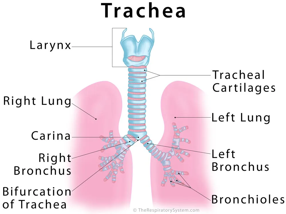
The trachea is located in the midline of the body, starting in the lower neck just below the larynx and ending in the upper chest behind the breastbone, where it divides into the right and left main bronchi. It begins at the lower edge of the cricoid cartilage, at about the level of the sixth cervical vertebra (C6), and ends at about the level of the sternal angle, between the fourth and fifth thoracic vertebrae (T4-T5).
Neck part and chest part
Anatomists divide the trachea into a cervical (neck) part and a thoracic (chest) part. The cervical part lies just under the skin of the front of the neck, which is why the rings can be felt above the notch at the top of the breastbone. The thoracic part passes through the superior mediastinum, the upper part of the central compartment of the chest between the two lungs. In a living person who is standing and breathing in deeply, the lower end can drop as low as the sixth thoracic vertebra.
What lies around the trachea
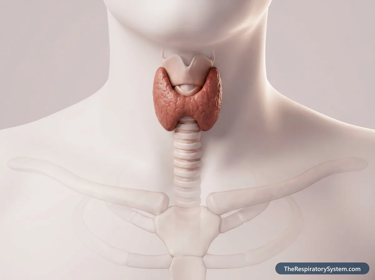
- In front: skin, fascia and the thin strap muscles of the neck; the isthmus of the thyroid gland, which crosses roughly the 2nd to 4th rings; lower down, the thymus (or its remnant in adults), the left brachiocephalic vein and the arch of the aorta.
- Behind: the esophagus, the muscular tube that carries food to the stomach, with the recurrent laryngeal nerves running in the grooves between the two tubes.
- On the sides in the neck: the lobes of the thyroid gland and the common carotid arteries.
- On the sides in the chest: the right vagus nerve, the arch of the azygos vein and the pleura on the right; the aortic arch, the left common carotid and left subclavian arteries and the left recurrent laryngeal nerve on the left.
Where is the trachea in relation to the esophagus?
The trachea lies directly in front of the esophagus along its whole length. Both tubes start near the same level in the neck, but the air passage is always the front one: food passes behind the windpipe, through the throat and down the esophagus. The epiglottis and the closing vocal folds keep swallowed food out of the trachea.
Where is the tracheal bifurcation?
The tracheal bifurcation, the point where the trachea splits in two, lies behind the sternal angle, the small ridge where the handle of the breastbone joins its body, roughly level with the second rib cartilages. Because the aortic arch presses on its left side, the lower trachea is pushed very slightly to the right of the midline.
Trachea Anatomy: Cartilage Rings, Trachealis Muscle and Carina
The trachea is built from 16 to 20 C-shaped rings of hyaline cartilage stacked one above another, joined by fibroelastic ligaments, and closed at the back by a membranous wall containing the trachealis muscle; at its lower end a ridge called the carina marks the split into the two main bronchi.
Where is the tracheal cartilage?
The tracheal cartilages lie in the wall of the tube, between the submucosa on the inside and the adventitia on the outside, and they cover the front and sides of the trachea. Each ring is about 1 to 2 mm thick and about 4 to 5 mm high, and the number varies from person to person. The rings are incomplete at the back, where the trachea touches the esophagus. The first ring is broader than the rest and is often joined to the next, and the last ring is thicker in the middle and curves down into the carina.
Between neighboring rings run the annular ligaments, bands of dense fibrous and elastic tissue. They let the trachea bend when the neck moves and lengthen during a breath in, the way the folds of a bellows let it stretch.
What is the trachealis muscle?
The trachealis muscle is a band of smooth muscle in the back wall of the trachea that spans the gap between the open ends of each cartilage ring. Most of its fibers run across the tube (transversely) and attach to the inner surface of the ring ends; a thinner outer layer runs lengthwise. Together with fibroelastic tissue it forms the membranous wall, the flat back of the windpipe that faces the esophagus. Because it is smooth muscle, it works without conscious control, under the autonomic nervous system.
What does the trachealis muscle do?
When the trachealis contracts, it pulls the two ends of each ring closer together and narrows the airway. A narrower tube makes air rush out faster during a cough, which helps blast mucus and particles up toward the throat. When it relaxes, the tube widens slightly, which lowers resistance to airflow during heavy breathing.
Carina of the trachea
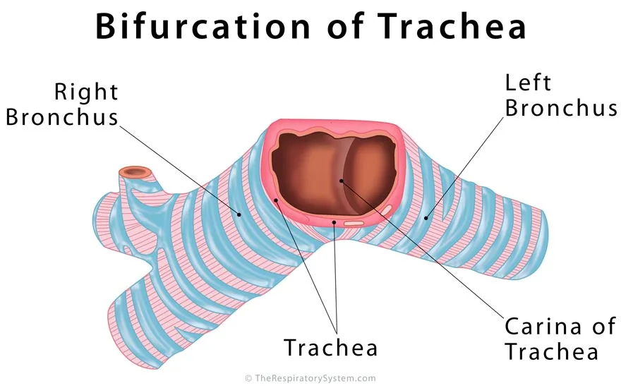
The carina is a keel-shaped ridge of cartilage at the bottom of the trachea, projecting upward between the openings of the right and left main bronchi. The lining over it is one of the most sensitive spots in the whole airway: touching it, or a bit of food landing on it, sets off a strong cough. Doctors looking down the airway through a bronchoscope use the carina as a landmark, and a widened or blunted carina can be a sign of enlarged lymph nodes below it.
Blood supply, nerves and lymph
The upper trachea receives blood from branches of the inferior thyroid arteries, and the lower part from the bronchial arteries; veins drain mainly into the inferior thyroid veins. Parasympathetic nerve fibers from the vagus nerve, many traveling in the recurrent laryngeal nerves, increase gland secretion and narrow the airway, while sympathetic fibers from the sympathetic trunk relax the smooth muscle. Lymph drains into the pretracheal and paratracheal lymph nodes and from there to the deep cervical and tracheobronchial nodes.
| Part | Where it is | What it does |
|---|---|---|
| Cervical trachea | Lower neck, from the cricoid cartilage (C6) to the top of the breastbone | Carries air from the larynx into the chest; easy to feel just above the sternal notch |
| Thoracic trachea | Superior mediastinum, behind the breastbone, down to the sternal angle (T4-T5) | Carries air to the bifurcation; widens slightly on each breath in |
| Tracheal cartilages | Front and sides of the wall, 16 to 20 C-shaped rings | Hold the airway open against changes in pressure |
| Annular ligaments | Between neighboring rings | Let the trachea bend and stretch with the neck and breathing |
| Trachealis muscle | Back wall, bridging the open ends of the rings | Narrows the tube during coughing and adjusts its diameter |
| Membranous wall | Flat back of the trachea, against the esophagus | Lets the esophagus bulge forward during swallowing |
| Respiratory mucosa | Innermost lining | Traps particles in mucus and sweeps them upward with cilia |
| Tracheal glands | Submucosa | Add mucus and watery fluid to the lining |
| Adventitia | Outermost layer | Anchors the trachea loosely to the esophagus and neck tissues |
| Carina | Inner ridge at the bifurcation, behind the sternal angle | Divides airflow into the two main bronchi; very sensitive trigger of coughing |
| Source: Typical adult anatomy, our editors | ||
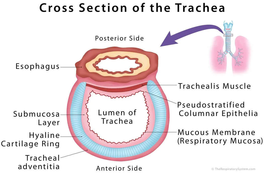
What Is the Trachea Made Of?
The trachea is made of four layers: an inner mucosa lined with ciliated pseudostratified columnar epithelium, a submucosa of loose connective tissue with glands, a layer of hyaline cartilage rings and trachealis muscle, and an outer adventitia of connective tissue. Cartilage is the material that makes up most of its bulk, and that is the reason it stays open: a rigid frame is needed so the tube does not collapse when air pressure inside it drops during a breath in.
Mucosa: what is the trachea lined with?
The inner surface of the trachea is lined with respiratory mucosa, made of pseudostratified ciliated columnar epithelium resting on a thick basement membrane. Most of the lining cells carry hundreds of hair-like cilia, and scattered among them are goblet cells that release mucus, basal cells that act as stem cells and replace worn-out cells, and a few brush cells and neuroendocrine cells. Under the epithelium lies the lamina propria, a thin layer of connective tissue rich in elastic fibers, small blood vessels and immune cells.
Submucosa
The submucosa is a layer of loose (areolar) connective tissue that contains collagen, elastin and reticular fibers, which keep the wall elastic. It holds the tracheal glands, small seromucous glands whose ducts open onto the surface and add watery fluid and mucus to the lining. The submucosa also carries the larger blood vessels, lymph vessels and nerves that serve the inner layers.
Cartilage and muscle layer
The third layer is the musculocartilaginous layer: the C-shaped hyaline cartilage rings at the front and sides, each wrapped in a fibrous perichondrium, and the trachealis muscle at the back. Hyaline cartilage is firm enough to resist collapse but bends slightly, so the trachea can flex with the neck and give way when a large mouthful of food passes down the esophagus behind it.
Adventitia
The outermost layer, the tracheal adventitia, is loose connective tissue that blends with the tissues around it. It anchors the trachea loosely to the esophagus, the thyroid gland and the other soft structures of the neck and mediastinum, while still letting the tube slide up and down during swallowing and breathing.
What Does the Trachea Do?
The trachea's main job is to conduct air between the larynx and the bronchi, and on the way it also warms, moistens and cleans that air and helps clear the airway by coughing. It is part of the conducting zone, the series of airways that move air but do not exchange gases. Our guide to respiratory system functions shows how this fits with the work of the rest of the system.
Function of the trachea in the respiratory system
On a breath in, air travels from the nasal cavity or the oral cavity through the pharynx and larynx, down the trachea and into the bronchi, which lead it into the lungs. On a breath out, air rich in carbon dioxide follows the same route in reverse. Because the trachea is the only route between the upper and lower airways, a blockage here affects both lungs at once.
Cleaning the air: the mucociliary escalator
The sticky mucus on the tracheal lining traps dust, pollen, smoke particles and microbes before they reach the lungs. The cilia under the mucus beat in coordinated waves, with a fast stroke toward the throat and a slow recovery stroke, so the mucus sheet moves steadily upward from the bronchi to the larynx. There it is coughed out or, much more often, swallowed without our noticing, and stomach acid destroys most of the trapped germs.
Warming and humidifying
Most warming and moistening of air happens in the nose, but the trachea finishes the job. By the time air passes the carina it is close to body temperature and nearly saturated with water vapor, which protects the delicate lining of the smaller airways from drying out.
The cough reflex
Nerve endings in the tracheal lining, and especially over the carina, detect irritants and send signals through the vagus nerve to the brainstem, which triggers a cough. During the cough the vocal folds close, pressure builds in the chest, and then the folds open suddenly. The trachealis contracts and the back wall bulges inward, narrowing the tube so the escaping air moves faster and drags mucus and debris with it.
Does the trachea have a role in the digestive system?
The trachea is not part of the digestive system, but it is built to share space with it. Its soft back wall lies against the esophagus, so when a mouthful of food passes down, the esophagus can bulge forward into the gap between the ring ends instead of being squeezed. At the same time, the larynx rises and the epiglottis tips over its entrance, so food and drink are steered behind the windpipe and away from the airway.
How Does the Trachea Work During Breathing?
The trachea is a passive tube during breathing: the diaphragm and the intercostal muscles change the size of the chest, and the trachea stretches, widens and narrows slightly as air moves through it.
Breathing in
When the diaphragm contracts and moves down, it pulls the lungs and the bronchi downward, and the trachea lengthens a little as the carina moves toward the abdomen. The thoracic part of the trachea widens slightly, because the pressure around it in the chest drops along with the pressure in the lungs. The cartilage rings keep it from being sucked shut by the fast inflow of air.
Breathing out
On a quiet breath out the trachea returns to its resting length and width. During a forced breath out or a cough, pressure in the chest rises above the pressure inside the airway, so the soft back wall of the thoracic trachea bows inward and the tube narrows. Healthy cartilage limits this narrowing; when the cartilage or the back wall is weak, the airway can close too much, which is the problem in tracheomalacia.
Air that never reaches the alveoli
The trachea, together with the nose, throat, larynx, bronchi and larger bronchioles, forms the anatomical dead space: about 150 mL of air in an adult that fills the conducting airways at the end of each breath in but never reaches the alveoli. That is why slow, deep breaths refresh the alveoli more efficiently than fast, shallow ones of the same total volume.

Trachea vs Esophagus: What Is the Difference?
The trachea is the airway that carries air to the lungs and lies in front, while the esophagus is the food pipe that carries food and liquid to the stomach and lies directly behind it. The two tubes run side by side through the neck and upper chest, but they are built very differently for their different jobs.
The trachea must stay open all the time, so it has cartilage rings and a ciliated, mucus-covered lining. The esophagus is a muscular tube that is collapsed when empty and opens only when a bolus of food passes, so it has no cartilage at all and is lined with tough stratified squamous epithelium that resists scraping by food. The table below compares them point by point.
| Feature | Trachea (windpipe) | Esophagus (food pipe) |
|---|---|---|
| Body system | Respiratory system | Digestive system |
| What it carries | Air | Food, liquid and saliva |
| Position in the neck and chest | In front | Directly behind the trachea |
| Starts at | Lower edge of the larynx (cricoid cartilage) | Lower end of the pharynx, at about the same level |
| Ends at | Carina, where it splits into two main bronchi | Stomach, after passing through the diaphragm |
| Approximate adult length | 10 to 12 cm | About 25 cm |
| Support | 16 to 20 C-shaped hyaline cartilage rings | No cartilage; layers of muscle only |
| Shape when at rest | Always open | Flattened and closed until food passes |
| Lining | Ciliated pseudostratified columnar epithelium with goblet cells | Nonkeratinized stratified squamous epithelium |
| Muscle | Smooth trachealis muscle in the back wall | Skeletal muscle in the upper part, smooth muscle lower down |
| Source: Typical adult anatomy, our editors | ||
How Does the Trachea Develop and Change With Age?
The trachea develops in the fourth week of embryonic life from the respiratory (laryngotracheal) diverticulum, an outpouching of the front wall of the foregut, the same early tube that forms the esophagus.
Before birth
As the respiratory diverticulum grows downward, ridges on each side fuse into the tracheoesophageal septum, which divides the foregut into the trachea in front and the esophagus behind. The lining epithelium and the glands of the trachea come from endoderm, while the cartilage, smooth muscle and connective tissue come from the surrounding splanchnic mesoderm. If the septum does not form properly, an abnormal channel called a tracheoesophageal fistula can be left between the two tubes, often together with a blind-ending esophagus (esophageal atresia).
Babies and children
A newborn's trachea is short, narrow and soft: its cartilage is less rigid than an adult's, so it can narrow more during crying or coughing. Because the airway is so small, even a little swelling of its lining cuts down airflow far more than it would in an adult. The trachea lengthens and widens steadily as the neck and chest grow, reaching adult size in the late teens.
Older adults
With age, the tracheal cartilages often become partly calcified and stiffer, and calcified rings may show up clearly on a chest X-ray or CT scan. The cilia may clear mucus less efficiently, and the cough reflex can become weaker, which is one reason chest infections are more common in later life.
Common Conditions That Affect the Trachea
Conditions of the trachea include narrowing, softening, abnormal connections to the esophagus, infection, injury, tumors and blockage, and each one affects a different part of the tube. Our respiratory diseases section covers airway disorders in more depth, and anyone with breathing symptoms should see a doctor.
- Tracheal stenosis: an abnormal narrowing of the tracheal lumen. It causes breathlessness, coughing, a high-pitched noise on breathing (stridor) and repeated chest infections. It can be present at birth but is more often acquired, most commonly as scarring after a breathing tube has been left in place for a long time (prolonged intubation) or after a tracheostomy.
- Tracheomalacia: weakness or floppiness of the cartilage or back wall, so the trachea collapses when pressure around it rises, especially on breathing out or coughing. The congenital form often improves on its own as a child's cartilage firms up over the first years of life; an acquired form can develop in adults.
- Tracheoesophageal fistula: an abnormal connection between the trachea and the esophagus, most often present from birth. Swallowed milk, food or saliva can pass into the airway and lungs, causing choking, coughing and lung infections.
- Airway obstruction: a blockage of the trachea, which can be as simple as a piece of food going down the wrong way or can result from swelling, infection or a tumor. A complete blockage is a medical emergency.
- Tracheitis: infection and inflammation of the trachea. Bacterial tracheitis is most common in children, often after a viral cold, and causes a deep cough, fever, noisy and difficult breathing and, in severe cases, a bluish tint to the skin.
- Tracheal tumors: primary cancers of the trachea are rare; the most common types are squamous cell carcinoma, which is strongly linked to smoking, and adenoid cystic carcinoma. Symptoms can include wheezing, a dry cough, breathlessness, hoarseness, coughing up blood and repeated chest infections.
- Tracheal injury or rupture: a tear in the tracheal wall, usually from blunt or penetrating trauma such as a car crash or a sports injury, and occasionally as a complication of a medical procedure.
- Saber-sheath trachea: a change in shape seen on imaging, in which the chest part of the trachea becomes narrow from side to side and long from front to back. It is strongly associated with chronic obstructive pulmonary disease (COPD).
- Tracheal deviation: a shift of the trachea away from the midline. It is a sign rather than a disease, pointing to something pushing the trachea, such as a large goiter or collapsed lung under pressure, or pulling it, such as a scarred or collapsed lung.
For people whose trachea is badly damaged over a long segment, researchers are studying tracheal replacement and transplantation, but these remain experimental.
Key Facts
- The adult trachea is about 10 to 12 cm long and about 2 cm wide, running from the larynx at the C6 level to the carina at the T4-T5 level.
- Sixteen to twenty C-shaped rings of hyaline cartilage keep the trachea open; they are incomplete at the back, where it touches the esophagus.
- The trachealis muscle bridges the open ends of the rings and narrows the windpipe to make coughs more forceful.
- The trachea divides at the carina into the right and left main bronchi, one for each lung.
- Ciliated cells and mucus-making goblet cells form a mucociliary escalator that carries trapped dust and germs up toward the throat.
- The trachea always lies in front of the esophagus, so the airway is the front tube and the food pipe is the back one.
- The trachea is part of the anatomical dead space, about 150 mL of air that never reaches the alveoli.
Quick Quiz: Test What You Know About the Trachea
6 questions. Tap an answer to check it.
How many C-shaped cartilage rings does the adult trachea usually have?
The trachea has about 16 to 20 C-shaped rings of hyaline cartilage, and the exact number varies between people.
What closes the gap at the back of each tracheal ring?
The open ends of the rings are bridged by the smooth trachealis muscle and a fibroelastic membrane, forming the flat back wall.
What is the carina?
The carina is a keel-shaped ridge of cartilage at the tracheal bifurcation, between the openings of the right and left main bronchi.
Which type of epithelium lines the trachea?
The trachea is lined by pseudostratified ciliated columnar epithelium with goblet cells, the typical respiratory epithelium.
Which structure lies directly behind the trachea?
The esophagus runs directly behind the trachea, in contact with its soft back wall.
At about which level does the trachea divide into the main bronchi?
The tracheal bifurcation lies behind the sternal angle, at about the level of the fourth to fifth thoracic vertebrae.
Frequently Asked Questions
What does it mean when the trachea is midline?
"Trachea midline" is a phrase doctors write after examining the neck. It means the windpipe sits in its normal central position, directly above the notch at the top of the breastbone, rather than being pushed or pulled to one side. A trachea that is off center, called tracheal deviation, can point to a problem in the chest or neck, so its position is a quick part of a physical exam.
What is tracheal tugging?
Tracheal tugging is a downward jerk of the trachea, felt or seen in the notch above the breastbone, that happens in time with each breath in. It usually shows that a person is working very hard to breathe, as the strongly contracting breathing muscles pull on the chest. A related tug that keeps time with the heartbeat was described in people with an enlarged aortic arch. It is a sign doctors look for, not a condition in itself.
What is a trachea tube?
"Trachea tube" usually means one of two medical devices. An endotracheal tube is passed through the mouth or nose, between the vocal folds and into the trachea, so a machine can help a person breathe during surgery or critical illness. A tracheostomy tube is placed through a surgical opening in the front of the neck directly into the trachea, usually below the first few rings, for longer-term breathing support.
What is a tracheal shave?
Despite its name, a tracheal shave does not touch the trachea. It is a cosmetic surgery, medically called chondrolaryngoplasty, that reduces the size of the Adam's apple, which is the front of the thyroid cartilage of the larynx. The surgeon shaves down part of this cartilage through a small incision in the neck. The windpipe itself lies lower and is not altered by the procedure.
Can you feel your trachea?
Yes. If you place a finger in the soft notch just above the top of your breastbone and press gently, you can feel the firm, ridged tube of the trachea in the midline. Moving your finger up, you reach the larger, harder cricoid and thyroid cartilages of the larynx. When you swallow, you can feel the whole airway rise and fall slightly as the larynx moves.
What happens when food goes down the trachea?
When food or drink slips past the epiglottis and vocal folds into the trachea, people say it went down the wrong pipe. The sensitive lining immediately triggers a strong cough to push it back out, and in most cases this works within seconds. If a larger piece blocks the airway completely, the person cannot cough, speak or breathe, which is a choking emergency that needs immediate help.
What is the difference between the trachea and the bronchi?
The trachea is a single tube in the neck and upper chest that carries air from the larynx. At the carina it divides into the two main bronchi, which enter the lungs and keep branching into smaller bronchi and bronchioles. Both have cartilage and ciliated lining, but the trachea has C-shaped rings, while the bronchi inside the lungs are supported by irregular plates of cartilage.
Is the trachea part of the upper or lower respiratory tract?
The trachea is the first part of the lower respiratory tract. Anatomists usually draw the dividing line at the lower edge of the larynx, at the cricoid cartilage: the nose, nasal cavity, sinuses, pharynx and larynx form the upper tract, and everything from the trachea down, including the bronchi, bronchioles and alveoli, forms the lower tract. Some textbooks count the larynx as part of the lower tract instead.
Can a person live without a trachea?
No. The trachea is the only passage between the larynx and the two main bronchi, so every breath in and out must pass through it. A person cannot breathe normally without a working windpipe, and a complete blockage is a medical emergency. Replacing a long section of trachea is very difficult, and engineered or transplanted windpipes are still being studied rather than used routinely.
Keep Exploring


