At a Glance
Alveoli (singular: alveolus) are microscopic air sacs in the lungs, each about 0.2 mm across, where gas exchange takes place. An adult has about 300 to 500 million of them, giving a surface of roughly 70 square meters. Oxygen diffuses from the alveoli into the capillary blood across a barrier only about 0.5 micrometers thick, and carbon dioxide diffuses the other way.
- Location
- Inside the lungs, at the ends of the respiratory bronchioles and alveolar ducts
- Function
- Gas exchange: oxygen into the blood, carbon dioxide out
- Tissue
- Type I and type II pneumocytes, alveolar macrophages, capillaries and elastic fibers
- Size
- Each about 0.2 mm across; about 300-500 million in an adult
- Connects to
- Bronchioles and alveolar ducts; pulmonary capillaries
- Part of
- Respiratory zone of the lungs, lower respiratory tract
On this page
- What Are the Alveoli?
- Where Are the Alveoli Located?
- Alveoli Anatomy: What Is the Structure of an Alveolus?
- What Are the Alveoli Made Of?
- What Do the Alveoli Do in the Respiratory System?
- What Happens in the Alveoli During Gas Exchange?
- How Do the Alveoli Work During Breathing?
- How Do the Alveoli Develop and Change With Age?
- Common Conditions That Affect the Alveoli
- Key Facts
- Quiz
- FAQ
What Are the Alveoli?
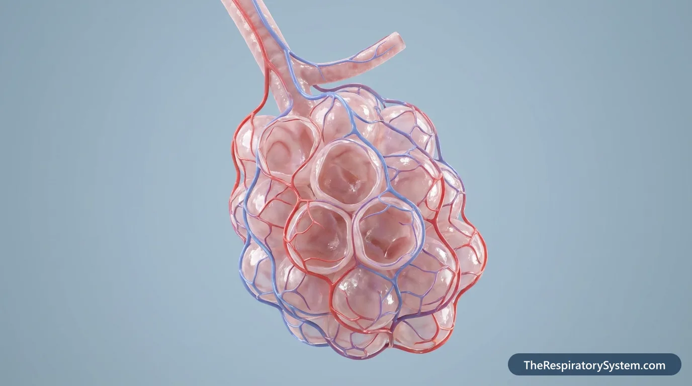
The alveoli are the microscopic air sacs of the lungs where the body takes in oxygen and gets rid of carbon dioxide, and they are the only part of the airway built for gas exchange. Air reaches them through the trachea, the bronchi and the bronchioles, and every alveolus is surrounded by blood capillaries. One sac is an alveolus; the plural is alveoli, pronounced al-VEE-uh-lie.
What do the alveoli look like?
Under a microscope, alveoli look like a mass of tiny, thin-walled bubbles or a bunch of grapes, packed so closely that neighboring sacs share their walls. Each alveolus is a cup-shaped pocket open on one side to an alveolar duct or sac. A typical alveolus is about 0.2 mm (200 micrometers) across, a little larger than the width of a human hair, and its size changes as the lungs fill and empty.
How many alveoli are in the lungs?
A healthy adult has about 300 to 500 million alveoli, roughly half in each lung, and the number is larger in people with bigger lungs. Because there are so many, their combined surface is huge, roughly 70 square meters, about the floor area of a small apartment or 40 times the area of the skin. This large surface is one of the main reasons gas exchange is so fast.
Explained simply
Picture hundreds of millions of tiny balloons at the ends of the lungs' smallest tubes. Each balloon is wrapped in a mesh of very thin blood vessels. When you breathe in, the balloons fill with fresh air; oxygen slips through their paper-thin walls into the blood, and carbon dioxide slips from the blood into the balloon to be breathed out.
Where Are the Alveoli Located?
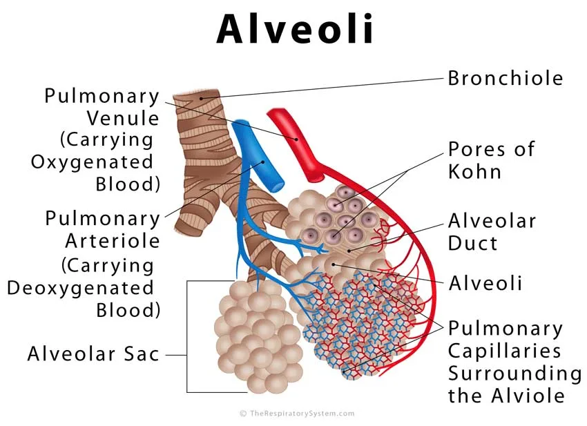
The alveoli are located inside the lungs at the very end of the bronchial tree: they bud from the walls of the respiratory bronchioles, line the alveolar ducts, and cluster at the ends of the ducts as alveolar sacs. They fill almost the whole volume of both lungs, in every lobe, from the apex up near the collarbone to the base on the diaphragm.
Where are the alveoli in the respiratory system?
Air on its way to the alveoli passes through the nose or mouth, the pharynx and the larynx, then down the trachea, through the main, lobar and segmental bronchi, and into the bronchioles. The last bronchioles, the respiratory bronchioles, divide into alveolar ducts, each leading to several alveolar sacs, groups of alveoli that open into a shared space. The cluster of respiratory bronchioles, ducts, sacs and alveoli beyond one terminal bronchiole is called a pulmonary acinus.
Are the alveoli connected to each other?
Yes. Neighboring alveoli share a single wall, the interalveolar septum, and many of these walls have small holes called pores of Kohn that let air pass from one alveolus to the next. As a result, the alveoli of a region form a connected, sponge-like network rather than a set of sealed bags.
Alveoli Anatomy: What Is the Structure of an Alveolus?
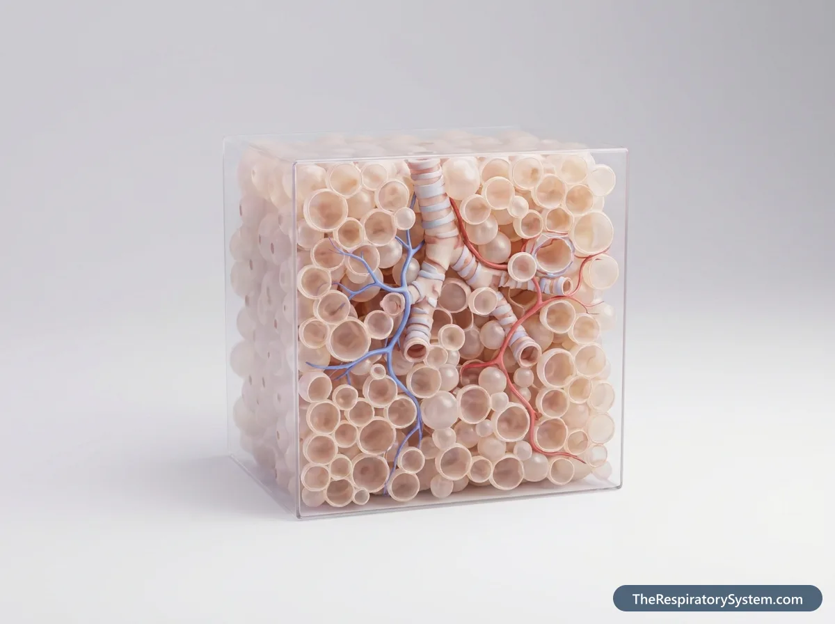
Each alveolus is a thin-walled sac made of a single layer of lining cells, surrounded by a dense mesh of capillaries held in a thin wall of connective tissue, with a film of surfactant on its inner surface. The wall between two alveoli, the interalveolar septum, is shared and contains the capillaries that serve both.
Interalveolar septum and interstitium
The septum is a thin partition with alveolar epithelium on both faces and a core of capillaries, elastic and collagen fibers, fibroblasts and a little tissue fluid. This core, part of the lung interstitium, holds the sacs in shape, lets each one stretch and recoil independently, and gives the lungs much of their elasticity. In healthy lungs the space is so thin that it is barely visible, but it thickens when it fills with fluid or scar tissue.
Pulmonary capillaries
The capillary network around the alveoli is so dense that blood flows around each sac almost like a thin sheet. Small branches of the pulmonary artery bring oxygen-poor blood into it, and pulmonary venules collect the oxygen-rich blood and lead it to the pulmonary veins. The capillaries are so narrow that red blood cells, about 7.5 micrometers across, pass through in single file and are squeezed against the wall, which shortens the distance oxygen has to travel.
Respiratory membrane
The respiratory membrane, also called the blood-air barrier, is the layer that gases cross. It is made of the thin type I alveolar cell, the capillary endothelial cell and their fused basement membranes, and in its thinnest parts it is only about 0.5 micrometers thick, less than a hundredth of the width of a hair. A thin film of fluid and surfactant covers it on the air side.
Pores of Kohn
Pores of Kohn are small openings, up to about 10 micrometers wide, in the walls between neighboring alveoli, bordered by fine elastic and reticular fibers. They allow collateral ventilation: if a small airway becomes blocked, air can still reach the alveoli beyond it through their neighbors. Alveolar macrophages can also move through them from one sac to the next.
| Part | What it is | What it does |
|---|---|---|
| Type I pneumocytes | Very thin, flat lining cells covering about 95% of the surface | Form the thin wall that gases cross |
| Type II pneumocytes | Rounded lining cells covering about 5% of the surface | Secrete surfactant and replace damaged type I cells |
| Alveolar macrophages | Wandering white blood cells on the inner surface | Engulf dust, debris and microbes |
| Surfactant layer | Thin film of phospholipids and proteins on the inner surface | Lowers surface tension so the alveoli do not collapse |
| Pulmonary capillaries | Dense network of tiny blood vessels in the alveolar walls | Bring oxygen-poor blood in and carry oxygen-rich blood away |
| Respiratory membrane | Alveolar cell, capillary cell and fused basement membranes | Barrier about 0.5 micrometers thick across which gases diffuse |
| Interalveolar septum | Shared wall between neighboring alveoli | Holds capillaries and elastic fibers; supports the sacs |
| Elastic and collagen fibers | Connective tissue fibers in the septum | Let alveoli stretch on a breath in and recoil on a breath out |
| Pores of Kohn | Small holes up to about 10 micrometers wide in the septum | Allow air to pass between neighboring alveoli |
| Source: Typical adult histology, our editors | ||
What Are the Alveoli Made Of?
The alveoli are made of two kinds of lining cells, flat type I pneumocytes and rounded type II pneumocytes, plus wandering alveolar macrophages, all resting on a thin basement membrane next to the capillaries. The inner surface is lined with a thin layer of fluid coated with surfactant.
Type I pneumocytes
Type I alveolar cells (type I pneumocytes, or squamous alveolar cells) are extremely thin, flat cells that spread out like fried eggs. Although they are fewer in number than type II cells, they cover about 95% of the alveolar surface, and their thinness makes diffusion of gases quick. They are delicate and cannot divide, so they are easily damaged by toxins or infection and must be replaced by type II cells.
Type II pneumocytes
Type II alveolar cells (type II pneumocytes, or great alveolar cells) are cube-shaped to rounded cells that often sit in the corners of the alveoli. They cover only about 5% of the surface, yet they outnumber type I cells, making up roughly 60% of the alveolar lining cells. They make, store and secrete pulmonary surfactant, and they act as stem cells: when type I cells are injured, type II cells divide and turn into new type I cells.
Alveolar macrophages
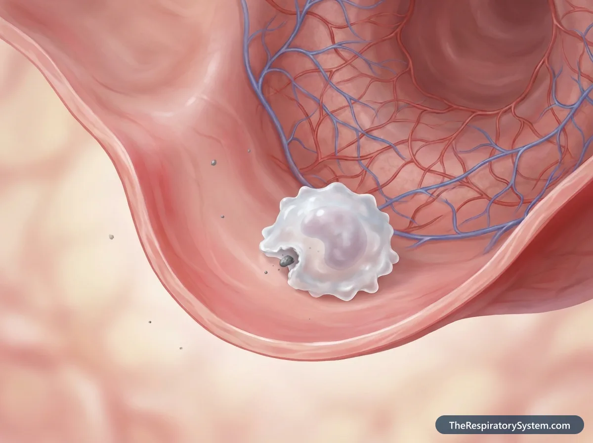
Alveolar macrophages, sometimes called dust cells, are white blood cells that crawl over the inner surface of the alveoli and through the walls between them. They engulf inhaled dust, soot, pollen and microbes that escaped the mucus and cilia of the larger airways, then either digest them or carry them up to the bronchioles to be cleared. This keeps the alveoli clean and helps prevent infection.
What are the alveoli lined with?
The alveoli are lined with simple squamous epithelium, made mostly of type I cells, which is in turn covered by a very thin film of watery fluid and a surface layer of surfactant. Surfactant is a mixture of about 90% lipids, mainly phospholipids, and about 10% proteins, which spreads across the air-liquid surface of every alveolus.
What Do the Alveoli Do in the Respiratory System?
The main function of the alveoli is gas exchange: they let oxygen from inhaled air pass into the blood and let carbon dioxide from the blood pass into the air to be breathed out. Every cell in the body needs a steady supply of oxygen to release energy from food, and produces carbon dioxide as waste, and the alveoli are where both gases cross between the air and the bloodstream. Our page on respiratory system functions shows how this fits into the work of the whole system.
Functions of the alveoli
- Taking in oxygen: oxygen diffuses from the air in the alveoli into the capillary blood and binds to hemoglobin in red blood cells.
- Removing carbon dioxide: carbon dioxide made by the body's cells diffuses from the blood into the alveoli and leaves with the next breath out.
- Helping control blood pH: because carbon dioxide forms an acid in the blood, the rate at which the alveoli unload it helps keep blood pH in a narrow, healthy range.
- Making surfactant: type II cells secrete surfactant, which keeps the alveoli from collapsing and makes the lungs easier to inflate.
- Defense: alveolar macrophages remove fine particles and microbes from the deepest parts of the lung.
- Repair: type II cells divide to replace damaged type I cells and restore the lining.
Function of the lungs and alveoli together
The lungs act as the frame and the bellows: the intercostal muscles and the diaphragm move air in and out, and the airways deliver it. The alveoli are the working surface at the end of that system, where the exchange of gases actually happens. Every other part of the respiratory system exists to bring air to the alveoli and blood past them.
What Happens in the Alveoli During Gas Exchange?
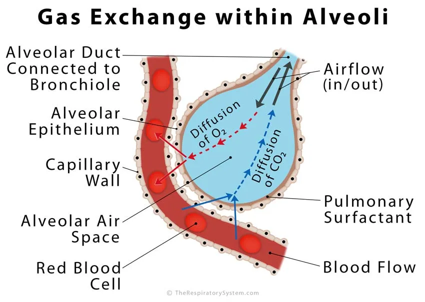
During gas exchange, oxygen moves by simple diffusion from the alveolar air, where its partial pressure is high, into the capillary blood, where it is low, while carbon dioxide moves the opposite way, from the blood into the alveoli. No energy is used; each gas simply moves down its own pressure gradient.
Step by step
- A breath in brings fresh air down the airways into the alveoli, raising the oxygen level and lowering the carbon dioxide level of the alveolar air.
- Blood arriving from the pulmonary arteries is low in oxygen and high in carbon dioxide, because it has just returned from the body's tissues.
- Oxygen dissolves in the thin fluid lining the alveolus, crosses the respiratory membrane and the blood plasma, and enters the red blood cells, where it binds to hemoglobin.
- At the same time, carbon dioxide leaves the blood, crosses the membrane into the alveolus, and is breathed out.
- The oxygen-rich blood flows into the pulmonary veins and back to the left side of the heart, which pumps it around the body.
Why the alveoli are so good at gas exchange
The speed of diffusion depends on three things: the surface area, the thickness of the barrier and the difference in gas pressure across it. The alveoli score well on all three, with an enormous surface, a barrier only about half a micrometer thick, and a steady pressure difference kept up by breathing and blood flow. Carbon dioxide dissolves far more readily than oxygen, so it crosses the membrane about 20 times faster, even though its pressure difference is smaller.
How fast gas exchange happens
At rest, a red blood cell spends about three-quarters of a second in an alveolar capillary, yet it is almost fully loaded with oxygen within about the first third of that time. This spare time is a safety margin: during hard exercise, when blood rushes through faster, the blood still has time to pick up its full load of oxygen in healthy lungs.
| Measure | Typical value | Why it matters |
|---|---|---|
| Number of alveoli | About 300 to 500 million | Creates a huge surface for gas exchange |
| Diameter of one alveolus | About 0.2 mm (200 micrometers) | Small sacs give more surface for the same volume |
| Total alveolar surface area | Roughly 70 square meters | About 40 times the area of the skin |
| Thickness of the air-blood barrier | About 0.5 micrometers in thin areas | A short path makes diffusion fast |
| Red blood cell diameter | About 7.5 micrometers | Cells pass the capillaries in single file, close to the wall |
| Time a red cell spends in an alveolar capillary | About 0.75 seconds | Leaves a safety margin during exercise |
| Time to load oxygen | About 0.25 seconds | Blood is fully loaded early in its transit |
| Oxygen partial pressure: alveolar air vs blood arriving | About 100 mmHg vs 40 mmHg | Drives oxygen into the blood |
| Carbon dioxide partial pressure: blood arriving vs alveolar air | About 46 mmHg vs 40 mmHg | Drives carbon dioxide into the alveoli |
| Source: Typical adult values, our editors | ||
How Do the Alveoli Work During Breathing?
The alveoli inflate when the chest expands during a breath in and shrink as the lungs recoil during a breath out, and surfactant keeps them from collapsing completely at the end of each breath out.
Breathing in and out
When the diaphragm contracts and the ribs lift, the pressure inside the alveoli drops slightly below the air pressure outside the body, and air flows in until the alveoli are filled. At the end of the breath the breathing muscles relax, and the stretched elastic fibers in the alveolar walls spring back, pushing air out. Even after a full breath out, about 1 to 1.5 liters of air stays in the lungs as residual volume, so the alveoli never empty completely.
How surfactant keeps the alveoli open
The thin film of water lining each alveolus has surface tension, a pull between water molecules that tends to shrink the sac. By Laplace's law, the pressure needed to keep a bubble open rises as the bubble gets smaller, so without help, small alveoli would empty into larger ones and collapse. Surfactant lowers surface tension, and it lowers it most when an alveolus is small and its surfactant molecules are crowded together. This keeps alveoli of different sizes stable, makes the lungs much easier to inflate, and helps keep fluid from being drawn into the air spaces.
Alveolar dead space
Some alveoli receive air but little or no blood flow, for example when a blood vessel is blocked, so the air in them takes no part in gas exchange. This wasted volume is called alveolar dead space. In healthy people it is very small; added to the anatomical dead space of the conducting airways, it makes up the physiological dead space.
How Do the Alveoli Develop and Change With Age?
The alveoli are the last part of the lung to form: the first primitive air sacs appear in the final months of pregnancy, and true alveoli keep forming after birth through early childhood.
Before birth
During the saccular stage, late in pregnancy, the ends of the airways widen into thin-walled saccules, and capillaries press up against them. Type II cells start making surfactant during the second half of pregnancy, and production rises sharply in the last weeks. True alveoli begin to form in the alveolar stage, at about the end of pregnancy, when new walls grow into the saccules and divide them into many smaller sacs.
Birth and childhood
At birth the alveoli are filled with fluid, which is quickly cleared as the baby takes its first breaths and surfactant helps the air sacs stay open. A newborn has only a fraction of the adult number of alveoli. Most of the rest form during the first years of life, and the alveoli keep enlarging as the chest grows.
Older adults
With age, the alveoli and alveolar ducts slowly become wider and shallower, and the total gas-exchange surface shrinks. The alveolar walls lose some of their elastic fibers, so the lungs recoil less on a breath out. These changes are normal and are different from emphysema, in which alveolar walls are actually destroyed.
Common Conditions That Affect the Alveoli
Conditions of the alveoli include infection, collapse, destruction of their walls, scarring, fluid in the air spaces and problems with surfactant, and each one interferes with gas exchange in a different way. Our respiratory diseases section covers them in more depth, and anyone with breathing symptoms should see a doctor.
- Pneumonia: an infection, usually bacterial or viral and sometimes fungal, that inflames the alveoli and fills them with fluid, pus and inflammatory cells. Oxygen cannot easily reach the blood through the filled sacs, which causes cough, fever and difficulty breathing.
- Emphysema: one of the two main forms of chronic obstructive pulmonary disease (COPD). The walls between alveoli are destroyed, leaving fewer, larger air spaces with less surface area and less elastic recoil, so air becomes trapped and the lungs become overinflated. Smoking is the main cause.
- Atelectasis: partial or complete collapse of part of a lung because its alveoli deflate. It is common after surgery under general anesthesia and can also result from a blocked airway, pressure on the lung or a lack of surfactant. Small areas often reopen as normal deep breathing and coughing return.
- Pulmonary edema: fluid leaking from the capillaries into the alveoli, most often because of heart failure, which makes it hard for oxygen to cross into the blood.
- Acute respiratory distress syndrome (ARDS): severe, widespread injury to the alveolar walls, for example after sepsis or a serious lung infection, so the alveoli flood with protein-rich fluid.
- Neonatal respiratory distress syndrome: breathing difficulty in premature babies whose type II cells have not yet made enough surfactant, so the alveoli collapse with each breath out.
- Pulmonary fibrosis: scarring and thickening of the alveolar walls and the tissue around them, which makes the lungs stiff and slows diffusion. When no cause can be found it is called idiopathic pulmonary fibrosis; other cases follow exposure to dusts, certain medicines or autoimmune disease.
- Alveolitis: inflammation of the alveoli. Extrinsic allergic alveolitis, also called hypersensitivity pneumonitis, is an allergic reaction to inhaled dusts such as mold spores or bird proteins.
- Alveolar rupture: bursting of overstretched alveoli, for example under the high pressures of mechanical ventilation, which can let air escape into the chest (pneumothorax).
- Alveolar capillary dysplasia: a rare congenital malformation in which the capillaries around the alveoli fail to develop properly and the pulmonary veins are misaligned. It causes severe breathing failure within hours to weeks of birth and is very often fatal.
Key Facts
- Alveoli are microscopic air sacs, each about 0.2 mm across, at the ends of the bronchioles.
- An adult has about 300 to 500 million alveoli with a combined surface of roughly 70 square meters.
- Type I pneumocytes cover about 95% of the alveolar surface, while type II pneumocytes make surfactant and repair the lining.
- Oxygen and carbon dioxide cross a respiratory membrane only about 0.5 micrometers thick, by simple diffusion.
- Surfactant lowers surface tension so the alveoli do not collapse at the end of each breath out.
- Alveolar macrophages, or dust cells, clear particles and microbes from the deepest parts of the lungs.
- Pores of Kohn link neighboring alveoli, so air can reach sacs beyond a blocked airway.
Quick Quiz: Test What You Know About the Alveoli
6 questions. Tap an answer to check it.
About how many alveoli does an adult have?
Adult lungs contain about 300 to 500 million alveoli, roughly half in each lung.
Which cells cover about 95% of the alveolar surface?
Thin, flat type I pneumocytes cover most of the surface and form the barrier that gases cross.
What is the main job of type II pneumocytes?
Type II cells secrete surfactant and can divide to replace damaged type I cells.
By what process does oxygen move from the alveoli into the blood?
Oxygen moves by simple diffusion from high partial pressure in the alveoli to lower partial pressure in the blood.
What are the pores of Kohn?
Pores of Kohn are small holes in alveolar walls that let air pass between neighboring alveoli.
Which condition destroys the walls between alveoli?
In emphysema the alveolar walls break down, leaving fewer, larger air spaces with less surface area.
Frequently Asked Questions
What does alveoli mean?
Alveoli is the plural of alveolus, a Latin word meaning a small hollow or cavity. In the lungs it names the tiny air sacs where gas exchange takes place. The same word is used elsewhere in the body for other small cavities, such as the milk-producing sacs of the breast and the tooth sockets in the jaw, so the context tells you which alveoli are meant.
What are alveoli in the breast?
Alveoli in the breast are tiny sac-like glands that produce milk. They are lined with milk-secreting cells and wrapped in small muscle cells that squeeze milk out into the ducts during breastfeeding. Clusters of these sacs form lobules within the breast. They share only a name with the alveoli of the lungs, which are air sacs for gas exchange.
What is alveolitis seca?
Alveolitis seca, better known as dry socket, is a dental problem and has nothing to do with the lungs. It happens when the blood clot in a tooth socket (the alveolus of the jaw) is lost or fails to form after a tooth is pulled, leaving bone exposed and causing pain. The name reflects the word alveolus, which also means a tooth socket.
What are the alveoli surrounded by?
Each alveolus is surrounded by a dense network of pulmonary capillaries, the tiniest blood vessels, which wrap around it like a net. These capillaries sit in the thin walls between neighboring alveoli, along with elastic and collagen fibers and a little tissue fluid. On the inside, the alveolus is coated with a thin layer of fluid and surfactant.
What is the difference between bronchi and alveoli?
The bronchi are large airways that carry air from the trachea into the lungs; they have cartilage, glands and a thick wall, and they exchange no gases. The alveoli are microscopic air sacs at the far end of the airway, with walls only one thin cell layer thick, where oxygen and carbon dioxide cross between the air and the blood. Between the two lie the bronchioles.
Can damaged alveoli be repaired?
The alveolar lining can repair itself after mild injury, because type II pneumocytes divide and turn into new type I cells. Alveolar walls that have been destroyed, as in emphysema, or replaced by scar tissue, as in pulmonary fibrosis, do not grow back in adults. Research into regrowing alveoli is ongoing, but at present damage of this kind is considered permanent.
Why are alveoli so small?
Many small sacs have a much larger total surface than a few big ones holding the same volume of air, and more surface means faster gas exchange. Small sacs also keep the distance from the air to the capillary blood very short. The cost is that small, wet bubbles tend to collapse, which is why the lungs need surfactant to keep the alveoli open.
Do alveoli have cilia?
No. Alveoli have no cilia and no mucus-making goblet cells, because a mucus layer would slow down gas exchange. Instead, particles that reach the alveoli are cleared by alveolar macrophages, which engulf them and either digest them or carry them up to the ciliated bronchioles, where the mucociliary escalator takes over and moves them toward the throat.
How do smoking and air pollution affect the alveoli?
Tobacco smoke and fine air pollution particles reach the alveoli, where macrophages engulf them and release chemicals that can injure the alveolar walls over time. In long-term smokers this damage can destroy alveolar walls and lead to emphysema, a form of COPD. Smoke also weakens the macrophages that defend the alveoli, making lung infections such as pneumonia more likely.
Keep Exploring


