At a Glance
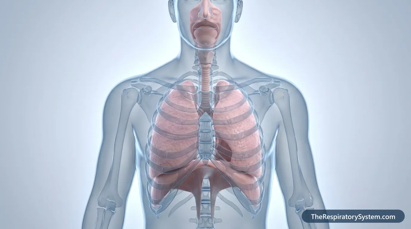
The respiratory system has three core functions: gas exchange, keeping blood pH in balance, and producing the airflow for speech. At rest an adult breathes about 12 to 20 times a minute, and across about 300 to 500 million alveoli in the lungs, oxygen enters the blood while carbon dioxide leaves it.
- Main function
- Gas exchange: oxygen into the blood, carbon dioxide out
- Other functions
- pH balance, cleaning and warming air, defense, speech, sense of smell
- Where gas exchange happens
- Alveoli of the lungs, across walls only one cell thick
- Breathing muscles
- Diaphragm and intercostal muscles
- Control center
- Medulla oblongata and pons in the brainstem
- Works with
- Circulatory, muscular, nervous, skeletal, digestive and urinary systems
On this page
- What Are the Main Functions of the Respiratory System?
- How Does Air Travel Through the Respiratory System?
- What Do the Nose, Mouth, Pharynx and Larynx Do?
- What Is the Function of the Trachea and Bronchi?
- What Is the Function of the Lungs and Alveoli?
- How Do the Diaphragm and Intercostal Muscles Help You Breathe?
- How Do the Respiratory and Circulatory Systems Work Together?
- How Does the Respiratory System Work With Other Body Systems?
- Key Facts
- Quiz
- FAQ
What Are the Main Functions of the Respiratory System?
The main function of the respiratory system is gas exchange, and around it sit several supporting jobs that keep the body's chemistry, defenses and voice working. Every cell needs a steady supply of oxygen (O2) to release energy from food, and every cell makes carbon dioxide (CO2) as waste. The body stores almost no oxygen, so a person cannot survive more than a few minutes without breathing, and the system keeps working day and night, awake or asleep.
The 8 main functions
- Gas exchange: oxygen moves from inhaled air into the blood, and carbon dioxide moves from the blood into the air that is breathed out.
- Ventilation: the breathing muscles move air in and out of the lungs so fresh air keeps reaching the alveoli.
- Acid-base balance (homeostasis): carbon dioxide forms carbonic acid in the blood, so breathing it out faster or slower keeps blood pH within a narrow range close to 7.4.
- Conditioning the air: the nose and airways filter, warm and moisten air before it reaches the delicate lung tissue.
- Defense: mucus, cilia, coughing, sneezing and immune cells trap and remove dust, pollen and microbes.
- Speech and sound: air pushed out of the lungs vibrates the vocal folds in the larynx.
- Sense of smell: sniffing carries odor molecules up to the olfactory area in the roof of the nasal cavity.
- Support jobs: filtering small clots and air bubbles out of the blood, activating the hormone angiotensin, helping blood return to the heart, and building pressure in the abdomen for coughing, lifting and pushing during childbirth.
How speech uses the airflow
Speech production starts with a breath in, which fills the lungs with the air that powers the voice. The second step is phonation: air pushed out through the larynx sets the vocal folds vibrating and makes a basic sound. The third step is articulation, in which the throat, mouth, tongue, lips, jaw and nose shape that sound into words.
Explained simply
Think of the body as a city of trillions of tiny factories, the cells. Each factory burns fuel with oxygen and puts out carbon dioxide as smoke. The respiratory system is the city's air supply and chimney at once: it hands fresh oxygen to the blood, which delivers it to every factory, and it gets rid of the smoke with every breath out.
How Does Air Travel Through the Respiratory System?
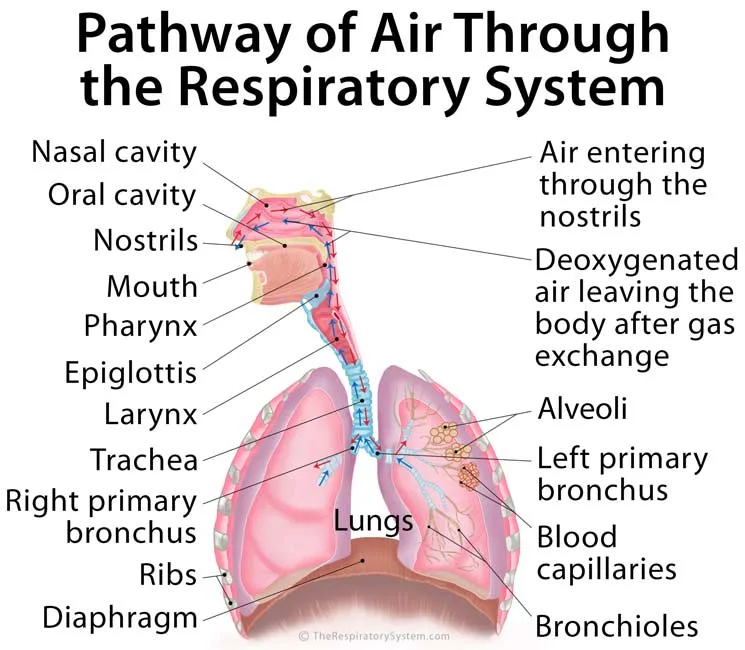
Air travels through the respiratory system along one fixed route: nose or mouth, pharynx, larynx, trachea, bronchi, bronchioles and finally the alveoli, where gas exchange takes place. The first part of this route, from the nostrils to the larynx, forms the upper respiratory tract. Everything from the trachea down to the alveoli forms the lower respiratory tract.
Conducting zone and respiratory zone
Anatomists also split the route by job. The conducting zone, from the nose to the terminal bronchioles, only moves, cleans and conditions the air; no gas exchange happens there, so the roughly 150 mL of air it holds is called anatomical dead space. The respiratory zone begins at the respiratory bronchioles, where the first alveoli bud from the airway walls, and continues through the alveolar ducts to the alveolar sacs.
Breathing out follows the same route in reverse. Because the airway is a dead-end tree rather than a one-way loop, each breath in mixes fresh air with some of the stale air left in the lungs. That is why the air inside the alveoli always holds less oxygen and more carbon dioxide than the air outside the body.
| Structure | Tract | What happens to the air |
|---|---|---|
| 1. Nostrils (nares) | Upper | Air enters; hairs catch the largest particles |
| 2. Nasal cavity | Upper | Air swirls over the conchae and is filtered, warmed and moistened |
| 3. Pharynx | Upper | Air passes the nasopharynx and oropharynx, a passage shared with food |
| 4. Larynx | Upper | Air passes between the vocal folds; the epiglottis guards the entrance during swallowing |
| 5. Trachea | Lower | Air is carried into the chest; mucus and cilia keep cleaning it |
| 6. Main bronchi | Lower | Air splits at the carina into the right and left lungs |
| 7. Bronchioles | Lower | Smooth muscle adjusts airflow down to the terminal bronchioles |
| 8. Respiratory bronchioles and alveolar ducts | Lower | The first alveoli appear and gas exchange begins |
| 9. Alveoli | Lower | Oxygen enters the capillary blood and carbon dioxide leaves it |
| Source: Typical adult anatomy, our editors | ||
What Do the Nose, Mouth, Pharynx and Larynx Do?
The nose, mouth, pharynx and larynx form the entrance of the airway: they let air in, clean and condition it, and steer it away from the food passage.
Function of the nose and nasal cavity
The nostrils are the main way in and out of the airway. Behind them, the nasal cavity is lined by a moist mucous membrane covered in cilia, tiny hair-like projections that beat toward the throat. Particles stick to the mucus and the cilia sweep them back to the pharynx, where they are swallowed, while larger particles are caught by hairs just inside the nose or blown out by sneezing. The scroll-shaped nasal conchae make the air swirl over this warm, wet surface, so by the time it reaches the throat it is close to body temperature and almost fully humidified.
The nose also works in reverse. On the way out, the cooler lining of the nasal cavity takes back part of the water vapor and heat from exhaled air, which limits water loss. The olfactory epithelium in the roof of the cavity carries the receptors for smell, and the air-filled paranasal sinuses that open into the nasal cavity lighten the skull and add resonance to the voice.
Function of the mouth
The oral cavity is the secondary entrance to the airway. It takes over when the nose is blocked and during hard exercise, when the body needs more air than the narrow nasal passages can deliver. The mouth is lined by a mucous membrane too, but it has no conchae and no ciliated respiratory lining, so it warms, moistens and filters air far less effectively than the nose.
Function of the pharynx
The pharynx, or throat, is a muscular tube behind the nasal cavity and mouth that passes inhaled air down to the larynx. Its upper part, the nasopharynx, carries only air, while the oropharynx and laryngopharynx carry both air and food. The tonsils and adenoids in its walls sample inhaled and swallowed germs as part of the immune system.
Function of the larynx
The larynx, or voice box, lets inhaled air pass into the trachea and exhaled air pass back toward the pharynx and nasal cavity. It holds the vocal folds, which vibrate to make sound, and it guards the lower airway. During swallowing the larynx rises and the leaf-shaped epiglottis folds back over its entrance while the vocal folds close, so food and drink are steered into the esophagus instead of the windpipe. If something slips past anyway, a forceful cough reflex usually throws it back out.
What Is the Function of the Trachea and Bronchi?
The function of the trachea (windpipe) and bronchi is to carry air between the larynx and the lungs while cleaning it and keeping the passage open. The trachea runs about 10 to 12 cm from the lower end of the larynx into the chest, where it divides into the two main bronchi at a ridge called the carina.
Function of the tracheal cartilage and trachealis muscle
About 16 to 20 C-shaped rings of hyaline cartilage keep the trachea from collapsing when the pressure inside it drops during a deep breath in. The rings are joined to one another by bands of fibrous and elastic tissue, which lets the windpipe bend and stretch as the neck moves. The open back of each C faces the esophagus and is closed by the trachealis, a band of smooth muscle. When it contracts, as in a cough, the trachea narrows and air rushes through faster, and its soft wall also lets the esophagus bulge forward when a large bite is swallowed.
How the windpipe cleans the air
The lining of the trachea and bronchi is pseudostratified ciliated columnar epithelium dotted with mucus-secreting goblet cells and backed by mucous glands. Mucus catches the dust and microbes that got past the nose, and the cilia beat upward in coordinated waves that carry the mucus to the throat. This conveyor-belt system is called the mucociliary escalator, and coughing speeds it up when extra mucus builds up.
Function of the bronchi and the bronchial tree
The right main bronchus carries air into the right lung and the left main bronchus into the left lung. Inside each lung they divide into lobar bronchi (three on the right, two on the left) and then segmental bronchi, the start of the branching bronchial tree. The walls of the trachea and bronchi contain smooth muscle, an involuntary muscle that the autonomic nervous system tightens or relaxes to regulate airflow. When the body needs more air, as during exercise, this muscle relaxes and the airways widen, while the cartilage keeps them from being squeezed shut.
Function of the bronchioles
The bronchioles are the small airways, about 1 mm wide or less, that carry air from the bronchi toward the alveoli. They have no cartilage, so their width depends on the smooth muscle in their walls, which can widen or narrow them to fine-tune where air goes. The last purely conducting branches are the terminal bronchioles; beyond them, respiratory bronchioles carry the first alveoli.
What Is the Function of the Lungs and Alveoli?
The function of the lungs is to keep a constant supply of oxygen flowing into the blood and to remove carbon dioxide from it, and the alveoli are where that exchange actually happens. The lungs keep air moving in and out so the gas inside the alveoli is renewed with every breath.
How the alveoli exchange gases
The alveoli are tiny air sacs clustered at the ends of the respiratory bronchioles and alveolar ducts, about 300 to 500 million in an adult. Each one is wrapped in a dense net of capillaries, and the barrier between air and blood is a single layer of flat cells on each side, in most places less than a thousandth of a millimeter thick. Oxygen diffuses across this barrier into the blood and carbon dioxide diffuses out, each gas moving from where its partial pressure is higher to where it is lower.
Pulmonary surfactant
Pulmonary surfactant is a mixture made mostly of phospholipids, with a few special proteins, that type II pneumocytes release onto the inner surface of the alveoli. It lowers the surface tension of the thin film of water lining each sac, so the alveoli, and with them the lungs, do not collapse at the end of a breath out. It also makes the lungs easier to inflate on the next breath in.
Lung lobes, pleura and the chest cavity
The right lung is divided into three lobes and the left lung into two, and all five lobes do the same job: they receive oxygen-poor blood from the body and send it back rich in oxygen. Both lungs sit in the thoracic (chest) cavity, enclosed by 12 pairs of ribs, the vertebral column and the sternum, which also house and shield the heart. Each lung is wrapped in pleura: the visceral pleura covers the lung surface, and the parietal pleura lines the inside of the chest wall. A thin film of pleural fluid between the two layers lets them glide without sticking and keeps the lung pressed to the chest wall, so it stays flexible and follows every breathing movement.
More jobs of the lungs
- Filtering the blood: the narrow pulmonary capillaries trap small clots and gas bubbles arriving in venous blood before they can reach the arteries of the body and cause an embolism.
- Hormone activation: angiotensin-converting enzyme on the lining of lung capillaries turns angiotensin I into angiotensin II, which helps regulate blood pressure.
- Excretion: carbon dioxide, water vapor and traces of volatile substances such as alcohol leave the body in exhaled air.
- Heat loss: warm, moist exhaled air carries away a small share of body heat.
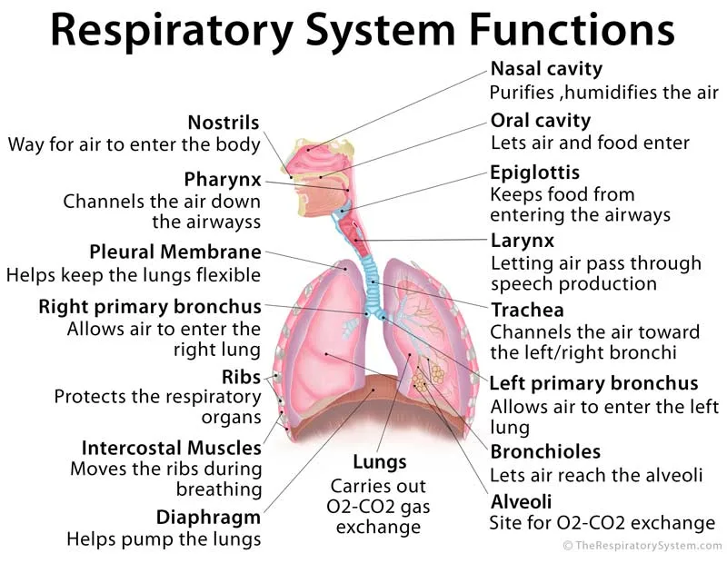
| Part | Main function | How it does it |
|---|---|---|
| Nose and nasal cavity | Filters, warms and humidifies air; smell | Hairs, mucus, cilia and the conchae; olfactory epithelium in the roof |
| Oral cavity | Secondary airway entrance | Takes over when the nose is blocked or during hard exercise |
| Pharynx | Channels air down to the larynx | Shared passage for air and food, with tonsils on guard |
| Epiglottis | Keeps food out of the airway | Folds over the larynx during swallowing |
| Larynx | Passes air; produces the voice | Vocal folds open for breathing and vibrate for speech |
| Trachea | Carries air into the chest | 16 to 20 C-shaped cartilage rings keep it open; cilia clear mucus |
| Bronchi | Carry air into each lung | Branch into the bronchial tree; smooth muscle regulates airflow |
| Bronchioles | Deliver air to the alveoli | Smooth muscle widens or narrows them |
| Alveoli | Gas exchange | Thin walls wrapped in capillaries; surfactant prevents collapse |
| Pleura | Lets the lungs move freely | Fluid film between two membranes |
| Diaphragm | Main breathing muscle | Contracts and flattens to enlarge the chest |
| Intercostal muscles | Move the ribs | Lift the rib cage on inhalation and lower it in forced exhalation |
| Source: Typical adult anatomy, our editors | ||
How Do the Diaphragm and Intercostal Muscles Help You Breathe?
The diaphragm and intercostal muscles help you breathe by changing the size of the chest cavity, because the lungs have no muscle of their own to inflate them. The lungs simply follow the chest wall: when the chest grows, the pressure inside the lungs falls and air flows in, and when the chest shrinks, air flows out.
Function of the diaphragm
The diaphragm is the primary muscle of respiration, a dome-shaped sheet of skeletal muscle just beneath the lungs that separates the chest cavity from the abdominal cavity. Its fibers attach to the lower ribs, the lower end of the sternum and the lumbar spine, and meet in a central tendon. When it contracts, the dome flattens and moves down, lengthening the chest and lowering the pressure in the lungs below the air pressure outside, so air rushes in. During quiet breathing the diaphragm does most of the work.
Function of the intercostal muscles
The intercostal muscles fill the 11 spaces between the ribs on each side in three layers: external, internal and innermost. The external intercostals lift the ribs up and out during inhalation, widening the chest from side to side and from front to back, while the internal intercostals pull the ribs down during forced exhalation. Together they also stiffen the chest wall so it is not sucked inward when the diaphragm pulls down.
Breathing in and breathing out, step by step
- Inhalation starts: the diaphragm contracts and flattens, and the external intercostals raise the rib cage.
- Air flows in: the chest cavity enlarges, the lungs stretch with it, and air flows in through the airways until the pressures are equal.
- Quiet exhalation: the muscles relax, the diaphragm returns to its dome shape, and the elastic lungs recoil, pushing air out without extra effort.
- Forced exhalation: during exercise, coughing or blowing, the internal intercostals and the abdominal muscles join in, pushing the abdominal organs up against the diaphragm.
Unlike the heart, these breathing muscles are skeletal muscles. Breathing runs automatically, even during sleep, but because these muscles are skeletal it can also be controlled on purpose, for example when holding the breath, singing or speaking.
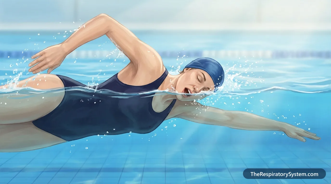
How Do the Respiratory and Circulatory Systems Work Together?
The respiratory and circulatory systems work together as one delivery network: the lungs load oxygen into the blood and unload carbon dioxide, and the heart and blood vessels carry both gases between the lungs and every cell. Together they also keep blood pH stable.
External respiration in the lungs
Oxygen-poor blood from the body returns through the superior and inferior venae cavae to the right atrium, passes into the right ventricle, and is pumped through the pulmonary arteries to the lungs. In the capillaries around the alveoli this blood has a high partial pressure of carbon dioxide and a low partial pressure of oxygen, while the fresh air in the alveoli has the opposite. Each gas diffuses down its own pressure gradient, oxygen into the blood and carbon dioxide into the alveoli, until the pressures nearly match. This exchange between the alveoli and the blood is called external respiration.
Internal respiration in the tissues
The newly oxygenated blood flows through the pulmonary veins to the left atrium and left ventricle, which pumps it through the aorta and its branches to the whole body. In the capillaries of the tissues the gradients are reversed: oxygen leaves the blood for the cells, and carbon dioxide from the cells enters the blood. This second exchange, between the blood and the body's cells, is called internal respiration. Inside the cells, the oxygen is then used in cellular respiration to release energy from food.
How the gases travel in the blood
Almost all the oxygen in blood is bound to hemoglobin in red blood cells, and only a small fraction dissolves in plasma. Carbon dioxide travels mostly as bicarbonate, with smaller amounts bound to hemoglobin or dissolved. The bicarbonate reaction links breathing to blood pH: when carbon dioxide builds up, more carbonic acid forms and the blood becomes more acidic, and faster breathing removes the excess.
Breathing also helps the circulation directly. Each breath in lowers the pressure in the chest and raises it in the abdomen, which helps draw venous blood back toward the heart, an effect called the respiratory pump.
How Does the Respiratory System Work With Other Body Systems?
The respiratory system also works with the nervous, skeletal, digestive and urinary systems: the nervous system sets the breathing rhythm, the skeleton protects and shapes the airway, and the digestive and urinary systems supply fuel and help balance blood chemistry.
Nervous system: what controls breathing?
Breathing is controlled by groups of neurons in the medulla oblongata, the lowest part of the brainstem, with help from centers in the pons. Chemoreceptors in the medulla and in the carotid and aortic bodies monitor carbon dioxide, oxygen and acid in the blood, and the strongest everyday trigger to breathe is a rise in carbon dioxide. The brainstem then adjusts the rate and depth of breathing, while nearby cardiovascular centers adjust the heart rate to match. Signals reach the two halves of the diaphragm through the left and right phrenic nerves, and reach the intercostal muscles through the intercostal nerves, which come from the thoracic spinal nerves T1 to T11.
Reflexes such as coughing, sneezing and yawning also run through the nervous system. The popular idea that people yawn because the body is short of oxygen has not held up in experiments, and the exact purpose of yawning is still debated. Smell links the two systems as well: olfactory nerve fibers from the nasal lining pass through the tiny holes of the cribriform plate of the ethmoid bone to reach the olfactory bulb, which rests on top of that plate.
Skeletal system
Bones and cartilages protect the soft respiratory organs and give the airway its shape. The perpendicular plate of the ethmoid bone, together with the vomer and the septal cartilage, forms the nasal septum that divides the nasal cavity into two sides, while the maxillae, palatine bones, nasal bones and conchae shape its walls and guide inhaled air in the right direction. The rib cage shields the lungs and heart from blows and moves with the breathing muscles, and the cartilages of the larynx, trachea and bronchi act as a flexible skeleton that holds the airway open.
Digestive and urinary systems
The digestive system supplies the nutrients that keep the cells of the airways and lungs alive, and the respiratory system supplies the oxygen that every cell of the digestive tract needs, with the blood carrying both. The pharynx is shared by the two systems, which is why swallowing and breathing must be tightly coordinated. The kidneys of the urinary system remove wastes and share the work of acid-base balance: the lungs adjust carbon dioxide within minutes, while the kidneys adjust acid and bicarbonate over hours to days. Problems in one system often show up in another, and our guide to respiratory diseases describes the conditions that affect breathing.
For a system-by-system tour, see how the respiratory system works with other body systems.
Key Facts
- Gas exchange happens only in the respiratory zone, from the respiratory bronchioles to the alveoli; the airways above hold about 150 mL of air that never reaches them, called dead space.
- A rise in carbon dioxide, not a fall in oxygen, is the main everyday signal that drives breathing.
- The diaphragm does most of the work of quiet breathing, and quiet breathing out is passive elastic recoil of the lungs.
- The nasal cavity warms and humidifies inhaled air so well that it is close to body temperature by the time it reaches the throat.
- By breathing out carbon dioxide, the lungs help hold blood pH close to 7.4, working together with the kidneys.
- About 16 to 20 C-shaped cartilage rings keep the trachea open, while the trachealis muscle closes the back of each ring.
- An adult at rest breathes about 12 to 20 times a minute, moving about half a liter of air with each quiet breath.
Quick Quiz: Test What You Know About the Functions
6 questions. Tap an answer to check it.
Where in the respiratory system does gas exchange take place?
Oxygen and carbon dioxide cross the thin walls of the alveoli and their capillaries. The trachea, bronchi and larynx only conduct air.
Which muscle is the primary muscle of breathing?
The diaphragm contracts and flattens to enlarge the chest, and it does most of the work during quiet breathing.
What does pulmonary surfactant do?
Surfactant, made by type II pneumocytes, reduces surface tension in the alveoli so they stay open at the end of a breath out.
Which part of the brain sets the basic rhythm of breathing?
Neuron groups in the medulla oblongata, helped by the pons, generate the breathing rhythm and adjust it to blood gas levels.
What happens to the blood when carbon dioxide builds up?
Carbon dioxide forms carbonic acid in the blood, so pH falls. Faster breathing removes the extra carbon dioxide.
Which structure folds over the entrance of the larynx during swallowing?
The epiglottis covers the laryngeal inlet so food and drink pass into the esophagus instead of the trachea.
Frequently Asked Questions
What is the function of the lungs for kids?
The lungs are like two big sponges inside your chest that fill up with air every time you breathe in. Their job is to take oxygen from that air and pass it into your blood, which carries it all around your body. At the same time, the lungs collect carbon dioxide, a waste gas your body makes, and send it out of your body when you breathe out.
What is the difference between breathing and respiration?
Breathing, also called ventilation, is the movement of air into and out of the lungs. Respiration is broader. External respiration is the gas exchange between the alveoli and the blood, internal respiration is the exchange between the blood and the body's cells, and cellular respiration is the chemical process inside cells that uses oxygen to release energy from food and produces carbon dioxide.
What is a lung function test?
A lung function test, or pulmonary function test, measures how much air the lungs hold, how fast air moves in and out, and how well oxygen passes into the blood. The most common is spirometry, in which a person breathes in as deeply as possible and then blows out hard and fast into a tube. Other tests measure total lung volume and gas transfer.
What are normal lung function test results?
Spirometry results are compared with predicted values for a healthy person of the same age, sex, height and ancestry. Values of the forced vital capacity (FVC) and the volume blown out in the first second (FEV1) at or above about 80 percent of predicted are usually treated as within the normal range. In adults, an FEV1 to FVC ratio below about 0.7 suggests narrowed airways.
Does the trachea have a function in the digestive system?
The trachea belongs to the respiratory system, not the digestive system, but the two work side by side. The windpipe lies directly in front of the esophagus, and its soft back wall, made of the trachealis muscle, lets the esophagus expand when food is swallowed. During swallowing the epiglottis and vocal folds close off the airway so food goes down the esophagus instead.
Why do we breathe faster during exercise?
Working muscles burn more fuel, so they use more oxygen and release more carbon dioxide and acid into the blood. Chemoreceptors and signals from the moving muscles and the brain itself tell the breathing centers in the brainstem to increase both the rate and the depth of breathing. The airways widen at the same time, and the heart pumps faster to carry the extra gases.
How many breaths does a person take in a day?
An adult at rest breathes about 12 to 20 times a minute, which works out to roughly 17,000 to 29,000 breaths a day, often rounded to about 20,000. The number rises with exercise, illness, fever or strong emotion and falls during deep sleep. Babies and young children breathe faster than adults, and their normal resting rate slows gradually as they grow.
How does the respiratory system change with age?
Lung function usually peaks in the early to mid-twenties and then declines slowly. Over the decades the lung tissue loses some of its elastic recoil, the rib cage stiffens, the breathing muscles weaken, and the cough and cilia become less effective at clearing mucus. Healthy older adults still breathe comfortably at rest, but they have less reserve during hard exercise or a chest infection.
Keep Exploring


