At a Glance
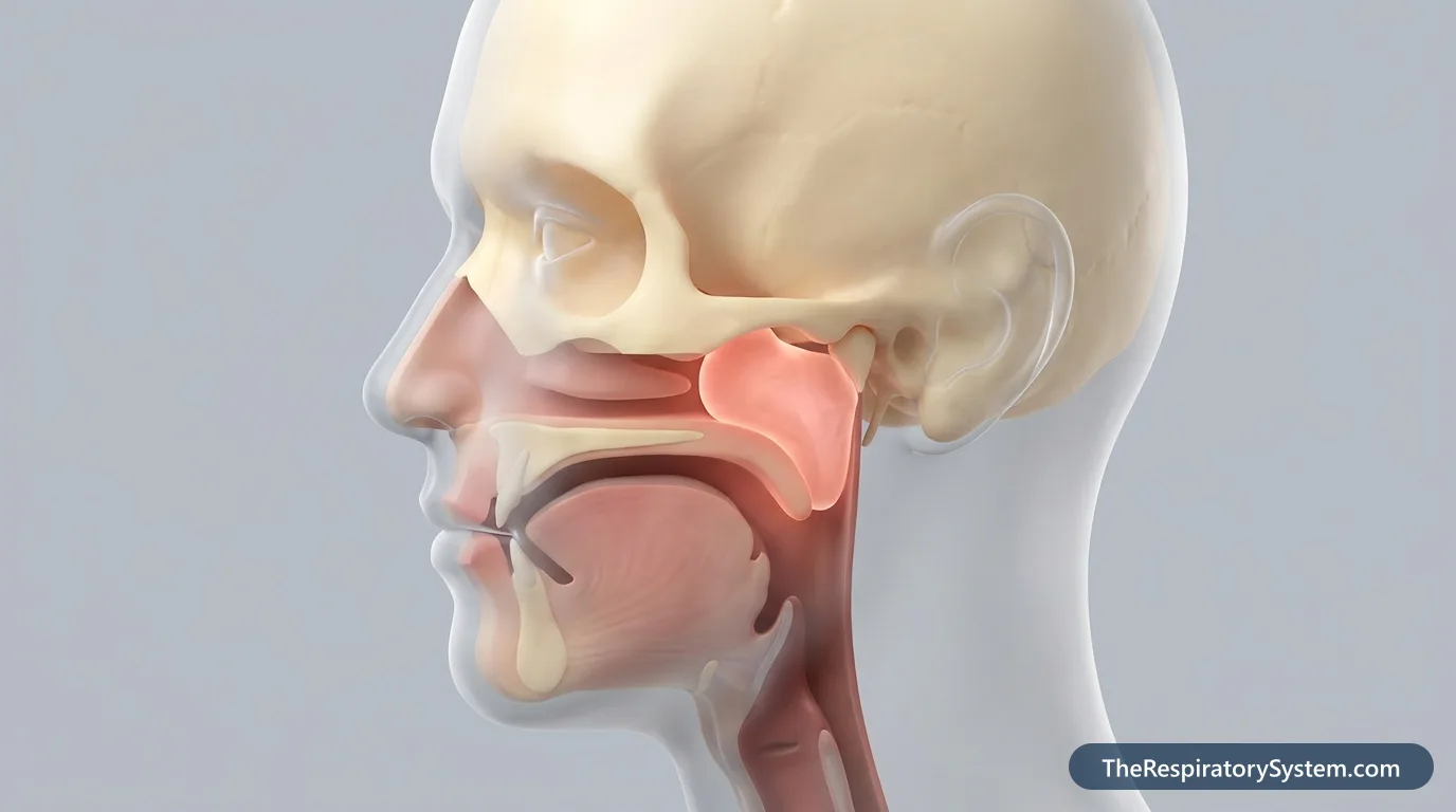
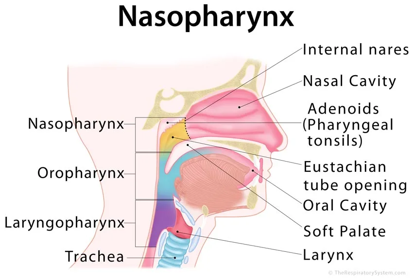
The nasopharynx is the top section of the throat, located behind the nasal cavity and above the soft palate. It starts at the choanae, the back openings of the nose, and ends at the level of the soft palate, where it continues as the oropharynx. It carries air from the nose toward the lungs, contains the adenoids and receives the two Eustachian tubes from the middle ears.
- Location
- Behind the nasal cavity, below the skull base, above the soft palate
- Function
- Air passage from the nose to the throat; immune defense; ventilates and drains the middle ear
- Tissue
- Respiratory (ciliated) epithelium and stratified squamous epithelium over lymphoid tissue and muscle
- Contains
- Adenoids (pharyngeal tonsil), Eustachian tube openings, torus tubarius, pharyngeal recesses
- Connects to
- Nasal cavity through the choanae; oropharynx below; middle ears through the Eustachian tubes
- Part of
- Pharynx, upper respiratory tract
On this page
What Is the Nasopharynx?
The nasopharynx is the first and highest of the three sections of the pharynx, the other two being the oropharynx and the laryngopharynx. It is the part of the throat that lies directly behind the nose, and it belongs to the upper respiratory tract. Its name combines naso, meaning nose, with pharynx, the Greek word for throat, and it is pronounced "nay-zo-FAIR-inks."
Unlike the lower parts of the throat, which carry both air and food, the nasopharynx is normally a passage for air only. Its walls are fixed to bone above and behind, so it stays open even when the muscles of the throat relax. During swallowing the soft palate rises and seals it off, which keeps food and drink from being pushed up into the nose.
Explained simply
Think of the nose as a hallway that ends at a back door. Behind that door is a small room, the nasopharynx, where air from both sides of the nose meets before it turns down into the throat. The room has a lump of germ-fighting tissue on its back wall (the adenoids) and a small tube on each side wall that leads to the ears.
Where Is the Nasopharynx Located?
The nasopharynx is located behind the nasal cavity and above the soft palate, in the hollow space just beneath the base of the skull and in front of the upper neck vertebrae. If you could look straight back through the nose past the nasal septum, the nasopharynx is the space you would reach.
Where does the nasopharynx start and end?
The nasopharynx starts at the choanae (also called the internal or posterior nares), the two openings at the back of the nasal cavity. It ends below at the level of the soft palate, where an opening called the pharyngeal isthmus leads into the oropharynx behind the oral cavity. From top to bottom it is only a few centimeters tall.
What is the nasopharynx posterior to?
- In front of it: the nasal cavity, separated only by the choanae and the back edge of the nasal septum, so the nasopharynx is posterior to the nasal cavity.
- Above and behind: the body of the sphenoid bone and the base of the occipital bone, which form a sloping roof and back wall.
- Behind: the first neck vertebrae and the deep muscles in front of them.
- Below: the upper surface of the soft palate, the boundary with the oropharynx.
- On each side: the openings of the Eustachian tubes and the deep pockets behind them.
Nasopharynx Anatomy: Walls, Openings and Adenoids
The nasopharynx is a small, box-shaped chamber with six boundaries: the choanae in front, a sloping roof and back wall against the skull base, the soft palate below, and two side walls that hold the openings of the Eustachian tubes. Several named landmarks sit on these walls.
Adenoids (pharyngeal tonsil)
The adenoids, also called the pharyngeal tonsil, are a mass of lymphoid tissue in the mucosa of the roof and back wall of the nasopharynx. Smaller clusters of lymphocytes are scattered under the lining elsewhere. The adenoids form the top of a ring of lymphoid tissue around the throat, together with the tubal tonsils near the Eustachian tube openings, the palatine tonsils in the oropharynx and the lingual tonsil at the back of the tongue.
Eustachian tube openings
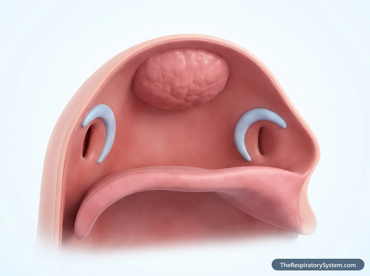
The pharyngeal openings of the Eustachian (auditory) tubes lie in the center of each side wall, roughly level with the lower edge of the inferior nasal concha. Each tube is a channel that runs back to the middle ear. Above and behind each opening, the cartilage of the tube raises a curved ridge called the torus tubarius, and a fold of mucosa, the salpingopharyngeal fold, runs down from it over the salpingopharyngeus muscle.
Pharyngeal recess
Behind each torus tubarius lies a deep, narrow pocket called the pharyngeal recess, or fossa of Rosenmüller. It is one of the hardest corners of the head and neck to see without an endoscope, which is why doctors examine it with a thin camera passed through the nose.
Muscles and fascia
The upper edge of the superior pharyngeal constrictor muscle does not reach the skull base. The gap is closed by a tough sheet of connective tissue, the pharyngobasilar fascia, which anchors the nasopharynx to the skull and keeps it rigid and open.
| Wall or landmark | What forms it | Notes |
|---|---|---|
| Anterior wall | Choanae and the back edge of the nasal septum | Open doorway to the nasal cavity |
| Roof | Body of the sphenoid bone | Slopes down into the back wall |
| Posterior wall | Basilar part of the occipital bone, upper neck vertebrae | Carries the adenoids |
| Floor | Upper surface of the soft palate | Rises to seal the nasopharynx during swallowing |
| Inferior opening | Pharyngeal isthmus | Leads into the oropharynx |
| Lateral walls | Mucosa over the pharyngobasilar fascia | Hold the Eustachian tube openings |
| Torus tubarius | Cartilage of the Eustachian tube | Ridge above and behind each tube opening |
| Salpingopharyngeal fold | Mucosa over the salpingopharyngeus muscle | Runs down from the torus tubarius |
| Pharyngeal recess | Deep pocket behind the torus tubarius | Also called the fossa of Rosenmüller |
| Source: Typical adult anatomy, our editors | ||
What Is the Nasopharynx Lined With?
The nasopharynx is lined mainly by two types of epithelium: stratified squamous epithelium, which covers about 60% of its surface in adults, and ciliated pseudostratified columnar respiratory epithelium with goblet cells, which covers most of the rest. It is the only part of the pharynx that has respiratory epithelium.
Histology of the nasopharynx
The respiratory epithelium is concentrated near the choanae and the roof, where it continues from the lining of the nose. The stratified squamous epithelium, which resists rubbing, is found mainly lower down near the soft palate and on the back wall. Where the two types meet, the border is usually sharp, but in some places a transitional, intermediate epithelium lies between them; respiratory and intermediate epithelium together make up the remaining 40% or so. Beneath the lining are mucous glands, lymphoid tissue and the muscles and fascia of the pharyngeal wall.
Blood supply and lymph drainage
Arterial blood comes from branches of the external carotid artery system: the ascending pharyngeal artery, the ascending palatine branch of the facial artery, and pharyngeal branches of the maxillary artery, including the artery of the pterygoid canal. Veins form a pharyngeal plexus that connects with the pterygoid plexus and drains toward the internal jugular vein. Lymph drains first to the retropharyngeal lymph nodes behind the pharynx and then to the deep cervical nodes of the neck.
What is the nasopharynx innervated by?
Sensation from the upper part of the nasopharynx is carried by the pharyngeal branch of the maxillary nerve (CN V2). The lower part receives sensory fibers from the glossopharyngeal nerve (CN IX) through the pharyngeal plexus. Most muscles of the area are supplied by the vagus nerve (CN X) through the same plexus, except the tensor veli palatini, which is supplied by the mandibular nerve.
What Does the Nasopharynx Do?
The nasopharynx carries air from the nasal cavity down to the rest of the airway, helps clean and condition that air, defends against inhaled germs through the adenoids, and connects the throat to the middle ears through the Eustachian tubes. It also helps shape the voice. These jobs are part of the broader respiratory system functions of the upper airway.
Function in the respiratory system
Every breath through the nose passes through the nasopharynx on its way to the oropharynx, the larynx, the trachea and the lungs. Its ciliated, mucus-covered lining continues the work of the nose, trapping particles and adding warmth and moisture before air moves further down. Mucus swept back from the nasal cavity and the paranasal sinuses collects here and is swallowed.
Immune defense
The adenoids and the scattered lymphoid tissue of the nasopharynx are among the first immune tissues to meet inhaled microbes. White blood cells circulating through them sample germs and particles and start immune responses against them, a role that is most active in childhood.
Ear drainage and pressure balance
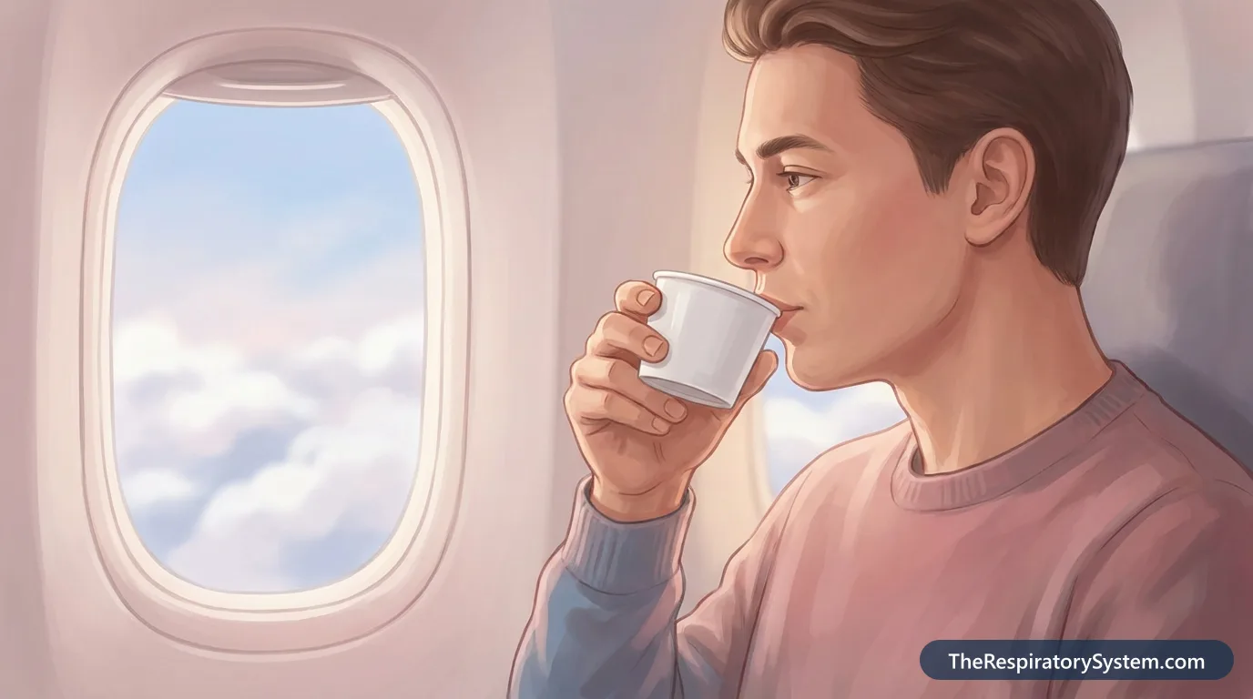
The Eustachian tubes link each middle ear to the nasopharynx. They open briefly during swallowing and yawning, letting air in to equalize pressure on both sides of the eardrum and letting secretions from the middle ear drain into the throat. The popping felt in the ears during a plane descent is this system at work.
Function in swallowing and speech
In the digestive system the nasopharynx carries no food, but it plays a part in swallowing: the soft palate rises against its back wall and closes it off so food does not enter the nose. In speech, it acts as a resonating chamber. Sounds such as m, n and ng need the nasopharynx to stay open so air and sound can pass into the nose, while other sounds need it closed.
Nasopharynx vs Oropharynx vs Laryngopharynx: What Is the Difference?
The nasopharynx, oropharynx and laryngopharynx are the upper, middle and lower parts of the same muscular tube, the pharynx; the nasopharynx carries only air, while the two lower parts carry both air and food. The table below compares them side by side.
The nasopharynx sits behind the nose and above the soft palate. The oropharynx lies behind the mouth, from the soft palate down to the level of the epiglottis, and it holds the palatine tonsils. The laryngopharynx, also called the hypopharynx, runs from the epiglottis down to the lower edge of the cricoid cartilage, where it continues as the esophagus behind the larynx.
Nasal cavity vs nasopharynx
The nasal cavity and the nasopharynx are neighbors, separated only by the choanae. The nasal cavity is divided in two by the septum and contains the conchae and meatuses, while the nasopharynx is a single shared chamber without conchae. Air from both sides of the nose merges in the nasopharynx.
How the nasopharynx changes with age
The nasopharynx develops from the upper part of the embryo's primitive pharynx, and the Eustachian tube grows from the first pharyngeal pouch. In babies the nasopharynx is small and the Eustachian tubes are shorter, wider and more horizontal than in adults, which helps explain why middle ear infections are common in early childhood. The adenoids grow during early childhood, are usually largest in the preschool and early school years, and then gradually shrink, so most adults have little adenoid tissue left.
| Feature | Nasopharynx | Oropharynx | Laryngopharynx |
|---|---|---|---|
| Position | Behind the nasal cavity | Behind the mouth | Behind the larynx |
| Upper limit | Skull base | Soft palate | Tip of the epiglottis |
| Lower limit | Soft palate | Tip of the epiglottis | Lower edge of the cricoid cartilage |
| Carries | Air only | Air and food | Air and food |
| Main lining | Respiratory and stratified squamous epithelium | Stratified squamous epithelium | Stratified squamous epithelium |
| Lymphoid tissue | Adenoids, tubal tonsils | Palatine and lingual tonsils | Scattered lymphoid tissue |
| Openings | Choanae, Eustachian tubes | Mouth (oropharyngeal isthmus) | Larynx in front, esophagus below |
| Source: Typical adult anatomy, our editors | |||
Common Conditions That Affect the Nasopharynx
Conditions of the nasopharynx mostly involve infection, enlarged adenoids, blocked Eustachian tubes or, rarely, tumors. Our respiratory diseases section covers airway conditions in more depth, and anyone with lasting symptoms should see a doctor.
- Nasopharyngitis (common cold): a viral infection that inflames the lining of the nose and nasopharynx, causing a runny or stuffy nose and a sore throat.
- Enlarged adenoids: large adenoids in children can partly block the back of the nose, leading to mouth breathing, snoring and a nasal-sounding voice, and they may block the Eustachian tube openings.
- Eustachian tube dysfunction and middle ear infections: swelling in the nasopharynx can stop the tubes from opening, trapping fluid behind the eardrum.
- Nasopharyngeal carcinoma: a rare cancer of the nasopharyngeal lining, often starting in the pharyngeal recess. Possible signs include a lump in the neck, a sore throat, a stuffy nose or nosebleeds, hearing loss with repeated ear infections, and blurred or double vision. It has been linked to Epstein-Barr virus infection, although most people with this very common virus never develop the cancer, and researchers are still studying genetic and other factors. It is much more common in parts of Southeast Asia and southern China than in the United States.
- Juvenile nasopharyngeal angiofibroma: a rare, noncancerous but blood-rich tumor near the back of the nose, seen mainly in adolescent boys.
Key Facts
- The nasopharynx is the uppermost of the three parts of the pharynx, above the oropharynx and laryngopharynx.
- It begins at the choanae behind the nasal cavity and ends at the level of the soft palate.
- It is the only part of the pharynx that normally carries air but not food.
- It is the only part of the pharynx lined in part by ciliated respiratory epithelium; stratified squamous epithelium covers about 60% of it in adults.
- The adenoids sit in its roof and back wall, and they usually shrink after childhood.
- The Eustachian tubes open into its side walls and balance the air pressure in the middle ears.
- Its sensory nerve supply comes from the maxillary nerve (CN V2) above and the glossopharyngeal nerve below.
Quick Quiz: Test What You Know About the Nasopharynx
6 questions. Tap an answer to check it.
Which part of the pharynx is the nasopharynx?
The nasopharynx is the top section of the pharynx, above the oropharynx and laryngopharynx.
What marks the lower boundary of the nasopharynx?
The soft palate separates the nasopharynx from the oropharynx below it.
Which structures open into the side walls of the nasopharynx?
Each Eustachian tube runs from the middle ear and opens into the center of a lateral nasopharyngeal wall.
Where are the adenoids located?
The adenoids (pharyngeal tonsil) lie in the mucosa of the roof and posterior wall of the nasopharynx.
Through which openings does air pass from the nasal cavity into the nasopharynx?
The choanae, or internal nares, are the back openings of the nasal cavity that lead into the nasopharynx.
What is the pharyngeal recess (fossa of Rosenmüller)?
The pharyngeal recess is a deep pocket behind each torus tubarius, a common starting site of nasopharyngeal carcinoma.
Frequently Asked Questions
What is a nasopharyngeal swab?
A nasopharyngeal swab is a thin, flexible swab passed through a nostril straight back along the floor of the nose until it reaches the back wall of the nasopharynx. It collects cells and mucus from that area, where many respiratory viruses multiply. The sample is then tested in a laboratory, for example for influenza, COVID-19 or other infections of the upper airway.
Why can the nasopharynx hurt?
Pain or soreness high in the back of the throat or behind the nose usually comes from inflammation of the nasopharyngeal lining, most often during a cold or other upper respiratory infection. Postnasal drip, dry air and swollen adenoids can also irritate the area. Because the nasopharynx is hidden behind the soft palate, it cannot be seen by simply opening the mouth.
Is the nasopharynx part of the digestive system?
Not really. The nasopharynx is part of the respiratory tract and normally carries only air. It does play a supporting role in swallowing, because the soft palate presses up against its back wall to close it off, which stops food and liquid from being pushed into the nose. The oropharynx and laryngopharynx below it are shared by the respiratory and digestive systems.
How do doctors look at the nasopharynx?
Because the nasopharynx sits behind the nose and above the soft palate, it is examined with a nasal endoscope, a thin tube with a light and camera passed through the nostril. Older methods used a small angled mirror placed at the back of the mouth. For deeper detail, doctors use imaging such as CT or MRI, which shows the walls, the adenoids and the surrounding skull base.
Where does the nasopharynx drain?
Mucus in the nasopharynx, including mucus swept back from the nose and sinuses, is carried down into the oropharynx and swallowed. Secretions from the middle ears drain into the nasopharynx through the Eustachian tubes. Lymph from its walls drains first to the retropharyngeal lymph nodes behind the throat and then to the deep cervical lymph nodes in the neck.
What does the nasopharynx look like?
Seen through an endoscope, the nasopharynx is a small, dome-shaped space with smooth pink lining. The back of the nasal septum divides the view at the front, the openings of the Eustachian tubes appear as slits on each side surrounded by a rounded ridge, and in children the bumpy, furrowed adenoids fill much of the roof and back wall.
What is the difference between the choanae and the nasopharynx?
The choanae are the two openings at the back of the nasal cavity, one on each side of the nasal septum. The nasopharynx is the single chamber that lies behind them. In other words, the choanae are the doorways, and the nasopharynx is the room that air enters after passing through them on its way down to the throat.
Do adults have adenoids?
Most adults have only a small amount of adenoid tissue left. The adenoids grow during early childhood, are usually largest in the preschool and early school years, and then gradually shrink during later childhood and adolescence. Enlarged adenoid tissue in an adult is unusual, so doctors look at it more closely when they find it.
Keep Exploring


