At a Glance
This glossary defines 31 respiratory terms in plain English. Six of them have their own in-depth pages: lung lobes, oral cavity, intercostal muscles, nostrils, choanae and nasopharynx. The other 25 are defined below from A to Z, each linked to the organ guide that explains it.
- Covers
- Anatomy and physiology terms of the upper and lower respiratory tract
- Entries
- 6 in-depth term pages plus 25 short A to Z definitions
- Level
- Grades 8 to 12 and introductory anatomy courses
- Start with
- The labeled respiratory system diagram, then the term you need
On this page
What Does This Respiratory Glossary Cover?
This glossary covers the structures of the airway and lungs, the muscles that move air, and the key words used to describe how gas exchange works. Terms are grouped from A to Z, and each one links to the page of our site that explains it in full, starting from the overview of the respiratory system.
Terms with their own in-depth pages
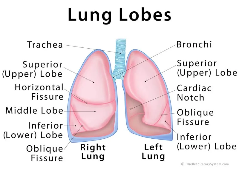
- Lung lobes are the separate sections of each lung, divided by deep grooves called fissures: three in the right lung and two in the left.
- Nostrils, or external nares, are the two outer openings of the nose, where air first enters the airway.
- Choanae, or internal nares, are the two back openings of the nasal passages, where the nasal cavity opens into the throat.
- The nasopharynx is the upper part of the pharynx, behind the nasal cavity, and the only part of the throat that carries air alone.
- The oral cavity, or mouth, is the secondary entrance to the respiratory tract, used when the nose is blocked or more air is needed.
- The intercostal muscles are the layers of muscle between the ribs that move the rib cage and are vital to breathing.
| Part | Where it is | What it does |
|---|---|---|
| Nostrils (nares) | Front of the nose | Let air in and out; hairs catch large particles |
| Nasal cavity | Behind the nose, above the roof of the mouth | Filters, warms and moistens air; holds smell receptors |
| Choanae | Back of the nasal cavity | Open the nasal cavity into the nasopharynx |
| Nasopharynx | Behind the nasal cavity, above the soft palate | Carries air down from the nose |
| Oral cavity | Mouth | Secondary entrance for air |
| Pharynx | Behind the nose and mouth | Carries air to the larynx and food to the esophagus |
| Larynx | Front of the neck | Guards the airway and produces the voice |
| Trachea | Neck and upper chest, in front of the esophagus | Carries air to the bronchi |
| Bronchi | Inside the chest, entering each lung | Carry air into the lungs and branch into the bronchial tree |
| Bronchioles | Inside the lungs | Carry air to the alveoli and adjust airflow |
| Alveoli | Ends of the smallest airways | Exchange oxygen and carbon dioxide with the blood |
| Lung lobes | 3 in the right lung, 2 in the left | Divide each lung into sections with their own airways and vessels |
| Diaphragm | Under the lungs, above the abdomen | Main breathing muscle |
| Intercostal muscles | Between the ribs | Move the rib cage during breathing |
| Source: Typical adult anatomy, our editors | ||
In-Depth Glossary Pages (20)


Respiratory System Terms A to F
Terms from A to F name the deepest parts of the airway, the reflex guards at its entrance and the main breathing muscle.
- Alveoli
- Tiny, thin-walled air sacs at the ends of the smallest airways, about 300 to 500 million in an adult, where oxygen enters the blood and carbon dioxide leaves it.
- Bronchi
- The two main airways that branch from the trachea into the right and left lungs, then divide into smaller lobar and segmental bronchi to form the bronchial tree.
- Bronchioles
- Small airways about 1 mm wide or less, with no cartilage in their walls, that carry air from the bronchi toward the alveoli.
- Carina
- The ridge of cartilage at the lower end of the trachea where it splits into the two main bronchi; its lining is very sensitive and triggers coughing.
- Cilia
- Microscopic hair-like projections on the cells lining the nasal cavity and airways that beat in waves to sweep mucus and trapped particles toward the throat.
- Diaphragm
- The dome-shaped sheet of muscle under the lungs that separates the chest from the abdomen; it contracts and flattens to draw air in and is the main muscle of breathing.
- Epiglottis
- A leaf-shaped flap of elastic cartilage at the top of the larynx that folds back during swallowing to keep food and drink out of the airway.
Respiratory System Terms G to N
Terms from G to N cover gas exchange itself, the voice box, the cleaning system of the airways and the parts of the nose.
- Gas exchange
- The movement of oxygen from the alveoli into the blood and of carbon dioxide from the blood into the alveoli by diffusion; it is the most important of the respiratory system functions.
- Glottis
- The vocal folds of the larynx together with the gap between them, called the rima glottidis; it opens for breathing and closes for swallowing and speaking.
- Goblet cells
- Cup-shaped cells in the lining of the nose, trachea and bronchi that release mucus to trap dust and microbes.
- Hilum
- The wedge-shaped doorway on the inner surface of each lung where the main bronchus, pulmonary vessels, nerves and lymph vessels enter and leave.
- Larynx
- The voice box: a tube of cartilages in the front of the neck that connects the pharynx to the trachea, protects the airway and holds the vocal folds.
- Lungs
- The two spongy organs in the chest where breathing ends in gas exchange; the right lung has three lobes and the left lung has two.
- Mucociliary escalator
- The cleaning system of the airways, in which mucus traps particles and cilia carry it up to the throat to be swallowed or coughed out.
- Nasal cavity
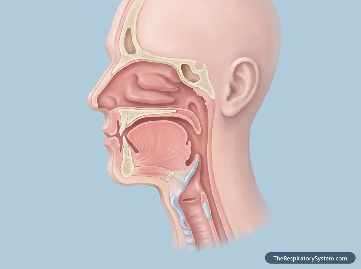
- The air-filled space behind the nose, divided in two by the nasal septum, that filters, warms and moistens inhaled air and holds the receptors for smell.
- Nasal conchae
- Three curled shelves of bone on each side wall of the nasal cavity, also called turbinates (superior, middle and inferior), that make inhaled air swirl over the moist lining.
- Nasal meatus
- Each of the air channels under a concha, the superior, middle and inferior meatus, into which the sinuses and the tear duct drain.
- Nose
- The external organ of the face made of bone and cartilage that houses the nostrils and forms the main entrance to the airway.
Respiratory System Terms P to Z
Terms from P to Z describe the air-filled spaces around the nose, the throat and windpipe, the lining of the lungs and how much air one breath moves.
- Paranasal sinuses
- Four pairs of air-filled spaces in the frontal, maxillary, ethmoid and sphenoid bones that open into the nasal cavity, lighten the skull and add resonance to the voice.
- Pharynx
- The throat: a muscular tube behind the nose and mouth, divided into the nasopharynx, oropharynx and laryngopharynx, that carries air to the larynx and food to the esophagus.
- Pleura
- The two-layered membrane around each lung; the visceral pleura covers the lung, the parietal pleura lines the chest wall, and a thin film of fluid between them lets the lung glide.
- Surfactant
- A mix of phospholipids and proteins made by type II pneumocytes that lowers surface tension inside the alveoli so they do not collapse when we breathe out.
- Tidal volume
- The amount of air moved in or out with one normal, quiet breath, about half a liter in an adult.
- Trachea
- The windpipe: a tube about 10 to 12 cm long, held open by 16 to 20 C-shaped rings of cartilage, that runs from the larynx to the main bronchi.
- Vocal folds
- Two bands of muscle and tissue inside the larynx, also called vocal cords, that vibrate as air passes between them to produce the voice.
How Does Air Move Through the Respiratory System?
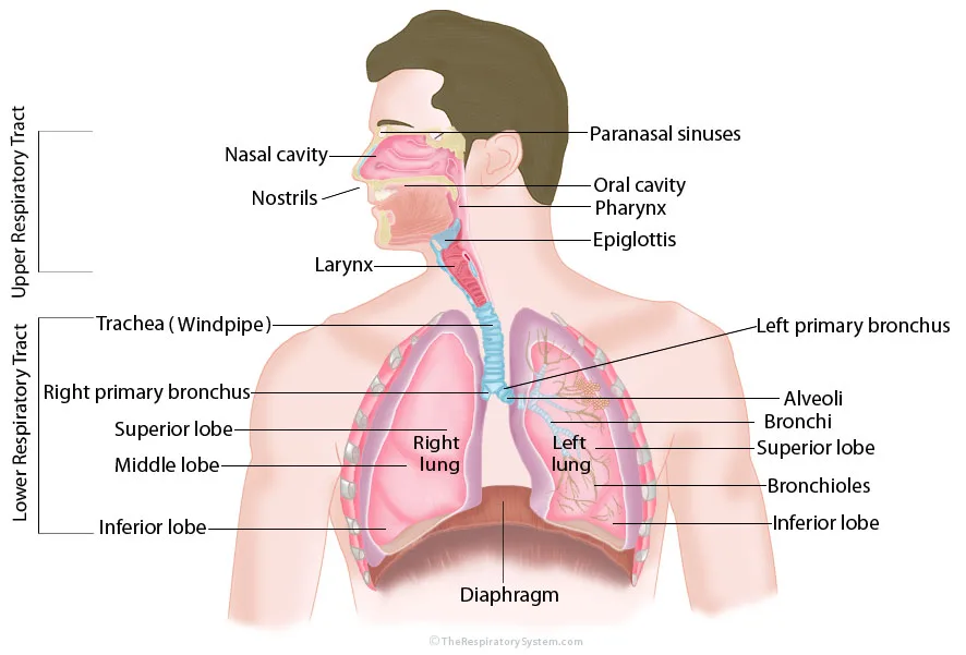
Air moves through the respiratory system in one line, from the nostrils to the alveoli, and the glossary terms follow that path in order. Every structure from the nostrils down to the larynx belongs to the upper respiratory tract, and every structure from the trachea down belongs to the lower respiratory tract.
- Air enters through the nostrils, or through the oral cavity when the nose is blocked.
- It flows through the nasal cavity, where the conchae and meatuses warm, moisten and filter it.
- It leaves the nasal cavity through the choanae and passes down the nasopharynx and the rest of the pharynx.
- It passes the epiglottis and the glottis of the larynx and enters the trachea.
- At the carina it divides into the two main bronchi, which branch into ever smaller bronchioles inside the lung lobes.
- It reaches the alveoli, where gas exchange takes place, while the diaphragm and intercostal muscles power each breath.
Explained simply
Picture a busy highway that starts at two front doors, the nostrils, and ends in millions of tiny parking spaces, the alveoli. Along the way, air is cleaned at checkpoints, warmed in the nasal cavity and sorted at junctions such as the carina. Most of the words in this glossary are simply names for the stops along that road.
Key Facts
- The choanae are the two back openings of the nasal cavity, where it joins the nasopharynx.
- The right lung has three lobes and the left lung has two.
- The intercostal muscles fill the spaces between the ribs in three layers: external, internal and innermost.
- The upper respiratory tract runs from the nostrils to the larynx, and the lower tract runs from the trachea to the alveoli.
- Gas exchange happens only in the alveoli and the smallest airways that carry them; every other part of the airway only conducts air.
- Many respiratory terms come from Greek and Latin: pneumon is Greek for lung, and pulmo is Latin for lung.
Frequently Asked Questions
What is the difference between the upper and lower respiratory tract?
The upper respiratory tract includes the nose, nasal cavity, paranasal sinuses, pharynx and larynx, the parts in the head and neck. The lower respiratory tract includes the trachea, bronchi, bronchioles and alveoli, which lie mostly inside the chest. Infections of the upper tract, such as colds, are usually milder than infections of the lower tract, such as pneumonia.
What is the smallest part of the respiratory system?
The smallest working units of the respiratory system are the alveoli, tiny air sacs only about 0.2 mm across. Their walls are made mostly of flat type I pneumocytes, and the barrier between the air inside them and the blood in the surrounding capillaries is, in most places, less than a thousandth of a millimeter thick. The smallest airways are the respiratory bronchioles and alveolar ducts that lead to them.
Why do respiratory terms come from Latin and Greek?
Anatomy took its vocabulary from the ancient Greek and Roman physicians and later from medieval and Renaissance scholars who wrote in Latin. That is why the lung is pulmo in Latin, giving the word pulmonary, and pneumon in Greek, giving pneumonia and pneumothorax. Naris is Latin for nostril, bronchus comes from the Greek for windpipe, and pleura is Greek for side or rib.
What is the difference between inhalation and exhalation?
Inhalation, or inspiration, is breathing in: the diaphragm and external intercostal muscles contract, the chest enlarges, and air flows into the lungs. Exhalation, or expiration, is breathing out. At rest it is passive, as the muscles relax and the elastic lungs recoil, but during exercise or coughing the internal intercostal and abdominal muscles help push the air out.
How many parts does the respiratory system have?
The count depends on how the parts are grouped, but the main structures are the nose and nasal cavity, the paranasal sinuses, the pharynx, the larynx, the trachea, the bronchi, the bronchioles, the alveoli and the two lungs that contain them. The diaphragm and intercostal muscles are usually included as the muscles of breathing, and the pleura wraps each lung.
What is the difference between the nasal cavity and the nasopharynx?
The nasal cavity is the air-filled space inside the nose, from the nostrils back to the choanae, lined with mucous membrane and shaped by the conchae. The nasopharynx begins right behind the choanae. It is the top part of the throat, above the soft palate, and it holds the adenoids and the openings of the auditory tubes that connect to the middle ears.
Keep Exploring


